Physical Therapy for Deformities of the Extremities
Quiz this lecture (99 questions)This lecture (Ahmed Omar & Nagwa Abuelwafa) walks joint by joint through the common deformities of the lower limb, then the ScapulaThe shoulder blade.General meaning, not from the slides, elbow and fingers. For each one it covers what the deformity is, what causes it (usually a pattern of tight vs lengthened/weak soft tissues or a bony MalalignmentBones or joints that are not lined up in their normal position.General meaning, not from the slides), and how physical therapy or surgery manages it.
A useful habit for this lecture: for almost every deformity, the cause list pairs a tight / shortened structure on one side with a stretched / elongated / weak structure on the other. Treatment follows the same logic: stretch the tight side, strengthen the weak side.
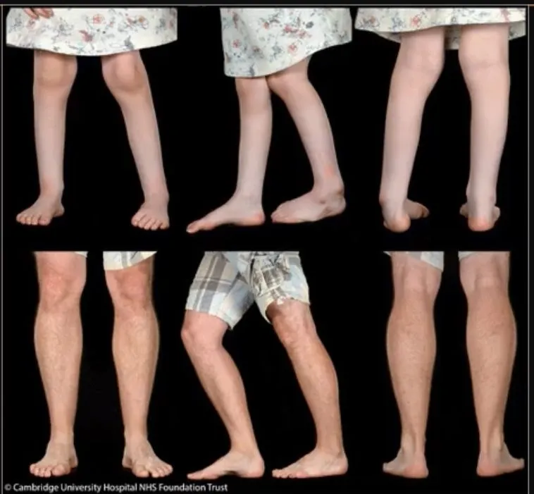
Objectives
| Verb | Objective |
|---|---|
| State | The physiotherapeutic approaches needed to solve various deformities |
| Describe | The principles of constructing a treatment plan and setting goals for ExtremityA limb: an arm or a leg, including the hand or foot.General meaning, not from the slides deformities, considering contextual factors |
| Explain | The physical, psychological and cultural needs of individuals with extremity deformities |
| Appraise | The suitable PTPhysical therapy (physiotherapy): treatment that uses exercise, movement and hands-on techniques to restore movement and function.General meaning, not from the slides assessment procedures for each case |
| Respect and consider | Patients’ differences, values, preferences and expressed needs in all professional activities |
Angles of the Femur
a) Neck–shaft angle (angle of inclination)
The angle in the Frontal planeThe vertical plane that divides the body into a front half and a back half.General meaning, not from the slides between the longitudinal axis of the Femoral neckThe narrower part of the femur between its rounded head and the shaft.General meaning, not from the slides and the longitudinal axis of the Femoral shaftThe long, straight middle part of the femur.General meaning, not from the slides.
- Normal adult: about 125°.
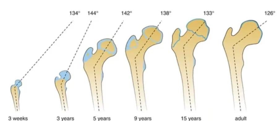
- Children: about 150°. Weight bearingPutting body weight through a limb or joint, for example by standing or walking on it.General meaning, not from the slides compresses the head and neck, so the angle decreases with growth (the slide’s growth figure: 134° at 3 weeks, 144° at 3 years, 142° at 5 years, 138° at 9 years, 133° at 15 years, 126° in adults).
Functions of the angle:
- It displaces the ProximalCloser to the trunk or the point of attachment of a limb.General meaning, not from the slides shaft laterally away from the joint, reducing the chance of bony ImpingementPinching or jamming of bone or soft tissue against another structure.General meaning, not from the slides against the pelvis.
- It allows more Degrees of freedomThe number of separate directions in which a joint can move.General meaning, not from the slides at the hip by moving the femoral axis away from the hip bone.
PathologicalCaused by disease or abnormal change, rather than being part of normal development.General meaning, not from the slides:
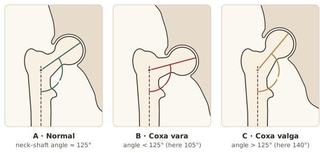
- Angle less than 125° → Coxa varaNeck–shaft angle of the femur below normal (less than 125°), so the femoral neck is bent inward toward the shaft. Means 'bend inward'. (“bend inward”), e.g. 105°.
- Angle more than 125° → Coxa valgaNeck–shaft angle of the femur above normal (above 125°; the coxa valga slide uses over 130°). Means 'bend outward'. (“bend outward”), e.g. 140°. (The coxa valga slide itself uses > 130° as the cut-off.)
b) Angle of torsion
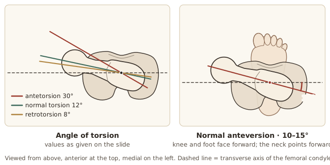
Viewed from above, the femoral neck’s longitudinal axis normally projects anterior to the medio-lateral (transverse) axis of the Femoral condylesThe two rounded knobs at the lower end of the femur that form the top of the knee joint.General meaning, not from the slides.
- Normally the angle faces medially and anteriorly at an average of 10–15° — this is Femoral anteversionThe forward twist of the femoral neck relative to the knee. Normally 10–15° in adults and about 30–35° in infants..
- In infants it is about 30–35°, because of the fetal position.
- Greater degrees of femoral anteversion or Femoral retroversionThe femoral head and neck point backward relative to the patellae, so the leg turns outward. may be seen.
Coxa Vara
Coxa varaNeck–shaft angle of the femur below normal (less than 125°), so the femoral neck is bent inward toward the shaft. Means 'bend inward'. is a progressive disorder of the ProximalCloser to the trunk or the point of attachment of a limb.General meaning, not from the slides end of the FemurThe thigh bone, the longest bone in the body.General meaning, not from the slides.
- Cause: unknown, but thought to be primarily an Endochondral ossificationThe process by which cartilage is gradually replaced by bone as bones grow.General meaning, not from the slides defect in the Femoral neckThe narrower part of the femur between its rounded head and the shaft.General meaning, not from the slides.
- IncidenceHow often a condition occurs in a population.General meaning, not from the slides: low, and equally distributed between the sexes.
Signs and symptoms
- Usually not recognised until the child begins to walk.
- A painless limp and the appearance of a short limb.
- May occur with excessive LordosisThe normal inward curve of the lower back. Excessive lordosis means the curve is exaggerated.General meaning, not from the slides, limited AbductionMoving a limb away from the midline of the body.General meaning, not from the slides, mild Hip flexion contractureA lasting shortening of the tissues at the front of the hip, so the hip cannot fully straighten.General meaning, not from the slides and limited hip extension.
Treatment of coxa vara
Goals:
- Increase the Neck–shaft angleThe angle, seen from the front, between the long axis of the femoral neck and the long axis of the femoral shaft..
- Promote OssificationThe process of bone forming and hardening.General meaning, not from the slides of the femoral defect.
- Reorient the Growth plateThe area of growing cartilage near the ends of a child's long bones, where lengthening happens.General meaning, not from the slides to a more horizontal position.
Decision by angle:
| Angle of inclination | Management |
|---|---|
| > 110° | Not treated, but carefully observed; a Heel liftA raised pad inside or under the heel of the shoe.General meaning, not from the slides, stretching of the Hip adductorsThe muscles on the inner thigh that pull the leg toward the midline.General meaning, not from the slides and the Lateral rotatorsThe muscles that turn the hip or leg outward.General meaning, not from the slides may be needed |
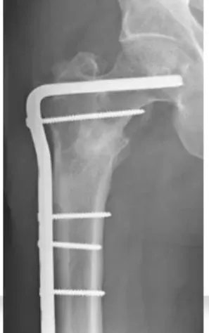
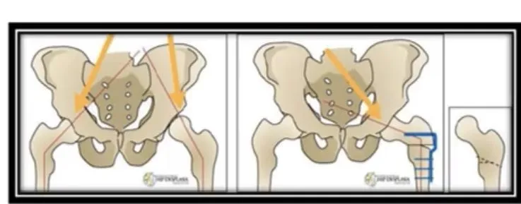
| < 110° | Surgery is the treatment of choice — a SubtrochantericJust below the trochanters, the bony bumps at the top of the femur.General meaning, not from the slides or IntertrochantericBetween the two trochanters, the bony bumps at the top of the femur.General meaning, not from the slides OsteotomyA surgical cut through a bone so that it can be realigned.General meaning, not from the slides fixed with a screw and plate or a Bifurcated blade plateA metal fixation device with a forked (bifurcated) blade and a plate, used to hold a cut bone in its new position.General meaning, not from the slides |
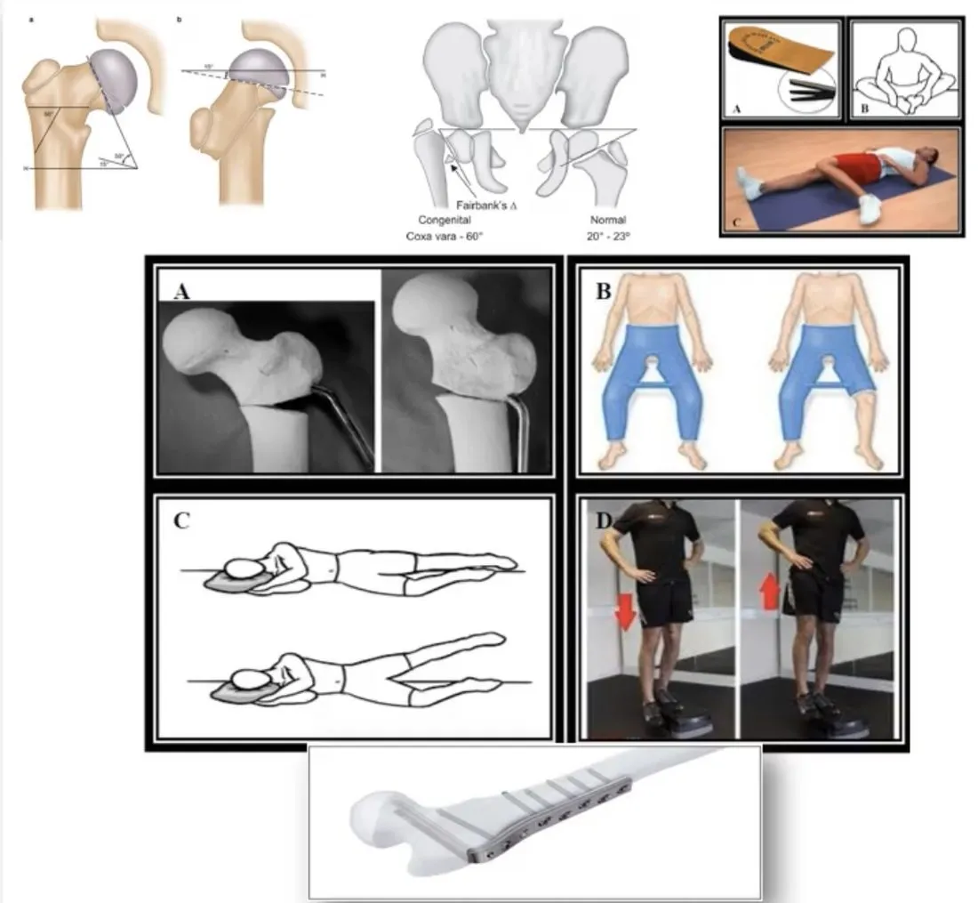
After surgery
- A Hip spica castA plaster cast that covers the trunk and part of one or both legs to hold the hip still.General meaning, not from the slides is applied for 6 to 8 weeks.
- After cast removal: active, gentle Range of motion (ROM)How far a joint can move in each direction.General meaning, not from the slides exercise for the hips and knee.
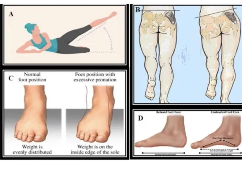
- Then active Side-lying hip abductionAn exercise lying on one side and lifting the top leg upward and away from the other leg.General meaning, not from the slides and standing Hip hikingLifting one side of the pelvis upward while standing. The notes call these Trendelenburg's exercises.General meaning, not from the slides (Trendelenburg’s) exercises.
- When full ROM is obtained: Partial weight bearingPutting only part of the body weight through the limb, usually with crutches.General meaning, not from the slides, Three-point crutch walkingA walking pattern with crutches in which both crutches and the weaker leg move together, then the stronger leg steps through.General meaning, not from the slides.
- When the Trendelenburg signA sign of weak hip abductors: when standing on one leg, the pelvis on the unsupported side drops.General meaning, not from the slides is negative, gait progresses to Full weight bearingPutting the whole body weight through the limb, with no crutches needed.General meaning, not from the slides.
Bifurcated blade plate exception: the blade is stable, so a spica cast is not necessary and partial weight bearing with crutches can begin 4 to 5 days after surgery. The osteotomy heals in about 8 weeks, after which active ROM and strengthening begin.
Coxa Valga
Neck–shaft angleThe angle, seen from the front, between the long axis of the femoral neck and the long axis of the femoral shaft. greater than 130°.
Causes:
- Trauma
- Congenital dislocation of the hipA hip joint that is out of its socket from birth.General meaning, not from the slides
- Spastic paralysisLoss of voluntary movement with abnormally tight, stiff muscles, caused by damage to the nervous system.General meaning, not from the slides
Signs and symptoms
- With moderate UnilateralAffecting one side of the body only.General meaning, not from the slides involvement, a painless limp.
- A Leg-length discrepancyOne leg is longer than the other.General meaning, not from the slides with the longer leg on the involved side.
- Hip adductorsThe muscles on the inner thigh that pull the leg toward the midline.General meaning, not from the slides tightness with upward Pelvic obliquityOne side of the pelvis sits higher than the other.General meaning, not from the slides, and weakness of the Hip abductorsThe muscles on the outer hip that lift the leg out to the side.General meaning, not from the slides on the affected side.
- Altered pelvic mechanics may cause back pain or Sacroiliac joint dysfunctionPain or abnormal movement at the sacroiliac joint, where the sacrum meets the hip bone.General meaning, not from the slides.
- Not seen as a problem in very young children (their angle is wider normally). In adults it is a real concern: pain, loss of mobility, possible death of tissue within the hip joint, difficulty bearing weight, limping and muscle deformity of the affected leg.
Treatment of coxa valga
- Femoral osteotomyA surgical cut through the femur so that it can be realigned.General meaning, not from the slides to correct the leg-length difference and restore normal mechanics.
- Stretch the adductors on the involved side and strengthen the abductors of the same side — these are essential.
- Teach the patient to keep the pelvis level to eliminate the limp.
- Watch the position of the feet; if incorrect, give foot-drill exercises (e.g. Short-foot exerciseAn exercise that draws the ball of the foot toward the heel to lift the arch, without curling the toes.General meaning, not from the slides / foot-core contraction for excessive PronationIn the foot: rolling inward so the arch flattens. In the forearm: turning the palm downward.General meaning, not from the slides).
- General leg exercises, but not many exercises in standing at first.
- Balance exercises and free exercises of all kinds to improve posture.
- Treat complications such as back pain and sacroiliac dysfunction.
Torsional Deformities of the Hip
1. Persistent femoral anteversion
- The Femoral headThe ball-shaped top of the femur that fits into the hip socket.General meaning, not from the slides and neck point forward in the same direction as the PatellaThe kneecap.General meaning, not from the slides. This is normal in a newborn.
- As bones grow, the head and neck spiral and Femoral anteversionThe forward twist of the femoral neck relative to the knee. Normally 10–15° in adults and about 30–35° in infants. falls to a maximum of about 15° in adults. Any delay in this process gives Persistent femoral anteversionFemoral anteversion that fails to fall to the adult value as the child grows, so the head and neck still point forward with the patellae..
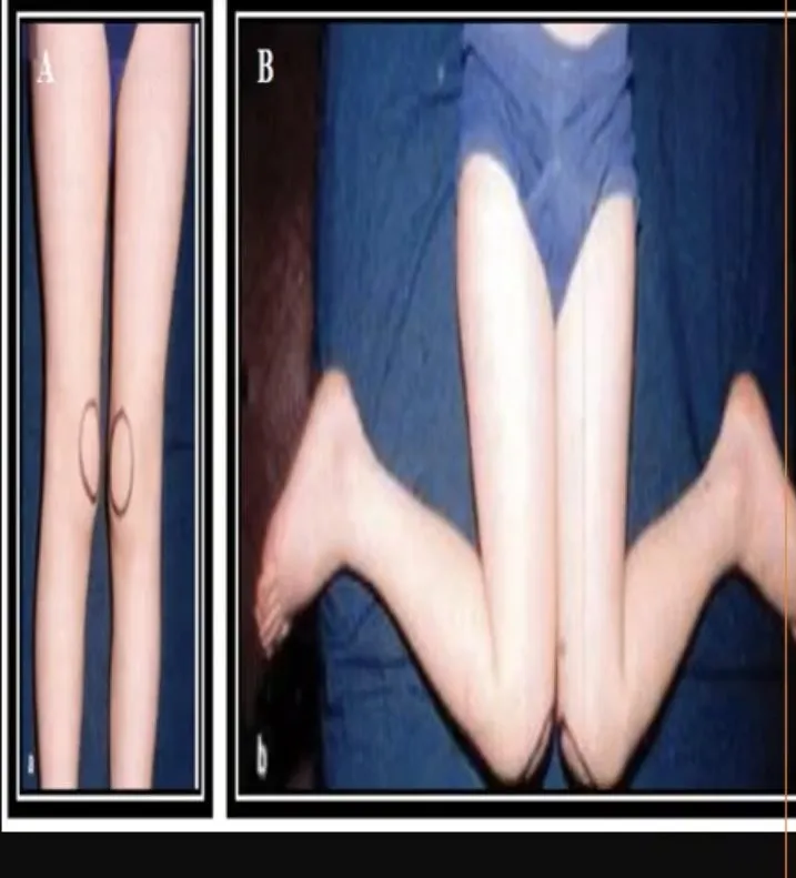
- The child must Internal rotationTurning a limb inward, toward the midline of the body.General meaning, not from the slides the FemurThe thigh bone, the longest bone in the body.General meaning, not from the slides to keep the femoral head in the AcetabulumThe cup-shaped socket in the pelvis that holds the head of the femur.General meaning, not from the slides → the whole leg turns in, with “Kissing patellaeKneecaps that face inward toward each other, because the whole leg is turned in.” and In-toeingWalking with the feet turned inward.General meaning, not from the slides. When running, the feet fly outwards.
- Children prefer to sit in the “W” position (feet spread laterally, knees bent, hips internally rotated).
- On examination: limited external rotation with legs extended, but internal rotation may exceed 90°.
- Conservative treatmentTreatment without surgery, such as exercise, stretching, bracing or casting.General meaning, not from the slides measures are not usually effective, but parents should encourage normal sitting.
- 80% resolve by age 8.
- If the cause is PathologicalCaused by disease or abnormal change, rather than being part of normal development.General meaning, not from the slides (e.g. Cerebral palsyA group of lifelong movement and posture disorders caused by damage to the developing brain.General meaning, not from the slides), anteversion worsens and Femoral osteotomyA surgical cut through the femur so that it can be realigned.General meaning, not from the slides between 8 and 10 years may be needed.
2. Femoral retroversion
- A rare cause of Out-toeingWalking with the feet turned outward.General meaning, not from the slides.
- Increased external rotation and decreased internal rotation at the hip.
- Associated with obesity; possibly with OsteoarthritisWear-and-tear arthritis in which joint cartilage thins and the joint becomes stiff and painful.General meaning, not from the slides, Stress fractureA small crack in a bone caused by repeated loading rather than a single injury.General meaning, not from the slides and SCFESlipped capital femoral epiphysis: the head of the femur slips off the neck at the growth plate.General meaning, not from the slides (slipped capital femoral epiphysis).
- The head and neck point backwards relative to the patellae, so the child External rotationTurning a limb outward, away from the midline of the body.General meaning, not from the slides the leg to seat the head — the “Charlie Chaplin stanceA stance with the legs turned outward so the patellae and feet point out, seen in femoral retroversion.” stance. Patellae and feet turn outward; the Medial longitudinal archThe arch along the inner side of the foot. The normal arch acts as a shock absorber. is reduced and the child looks flat-footed.
- Children with Dysplastic hipA hip whose joint has formed abnormally, for example with a shallow socket.General meaning, not from the slides occasionally present with Femoral retroversionThe femoral head and neck point backward relative to the patellae, so the leg turns outward., so always look for Hip instabilityA hip joint that is too loose or slips out of place too easily.General meaning, not from the slides.
- Rarely needs surgery — most resolve within a year of starting to walk.
3. Lateral rotation (patellae angle out)
May be due to:
- Tightness of the Lateral rotatorsThe muscles that turn the hip or leg outward.General meaning, not from the slides and Gluteus maximusThe large buttock muscle that extends and turns the hip outward.General meaning, not from the slides.
- Weakness of the Medial rotatorsThe muscles that turn the hip or leg inward.General meaning, not from the slides.
- Femoral retroversion.
- Internal tibial torsionThe feet face directly forward or inward (toeing in) because of a twist in the tibia. (CompensatedThe body adjusts another part to make up for a problem elsewhere.General meaning, not from the slides).
4. Medial rotation (patellae angle in)
May be due to:
- Tightness of the Iliotibial bandA thick band of connective tissue down the outer thigh, from the hip to just below the knee.General meaning, not from the slides and the medial rotators.
- Weakness of the lateral rotators.
- Femoral anteversion to a pathological degree (> 60–65°).
- External tibial torsionThe distal tibia is rotated laterally by more than the normal 25° from the proximal end, so the foot toes out. (compensated).
- Origin of in-toeing between hip and knee.
The most common in-toeing cause in children older than 3 years; associated with W-sittingSitting with the feet spread out to the sides, knees bent and hips turned inward, so the legs form a W. and an “Egg-beater running patternA running style in which the feet swing outward in circles, like an egg beater or windmill.General meaning, not from the slides” / “windmill” running pattern, and almost always corrects gradually by age 10.
Pelvis
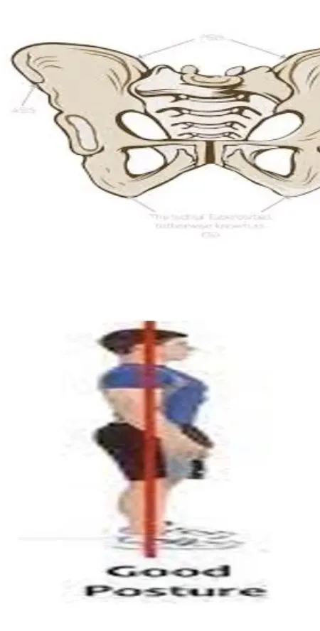
Normal alignment: the Plumb lineA string with a weight on the end, hung to give a true vertical reference line for judging posture.General meaning, not from the slides bisects the Gluteal cleftThe crease between the two buttocks.General meaning, not from the slides, the PSISPosterior superior iliac spine: the bony bump at the back of the pelvis, felt beside the base of the spine.General meaning, not from the slides are on the same horizontal plane, and the Iliac crestThe top curved edge of the hip bone, which you can feel at the waist.General meaning, not from the slides, Gluteal foldsThe skin creases under the buttocks, where the buttocks meet the thighs.General meaning, not from the slides and Greater trochanterThe bony bump at the top of the outer thigh, where several hip muscles attach.General meaning, not from the slides are level.
Common faults: Lateral pelvic tiltOne side of the pelvis is higher than the other. It is named for the side it tilts to., Anterior pelvic tiltThe pelvis tips forward: the superior pubic ramus lies inferior to the posterior inferior iliac spine., Posterior pelvic tiltThe pelvis tips backward: the superior pubic ramus lies superior to the posterior inferior iliac spines., Pelvic rotationThe pelvis is twisted so that the plumb line falls to the right or left of the gluteal cleft., Abducted hipA hip held out to the side, with the greater trochanter higher on the involved side..
1. Lateral pelvic tilt
One side of the pelvis is higher than the other. It is named for the side it tilts to — so a high right pelvis is called a left pelvic tilt.
Causes:
- ScoliosisA sideways curve of the spine.General meaning, not from the slides with IpsilateralOn the same side of the body as the structure being discussed.General meaning, not from the slides LumbarRelating to the lower back, the part of the spine between the chest and the pelvis.General meaning, not from the slides ConvexityThe outward-bulging side of a curve.General meaning, not from the slides
- Leg-length discrepancyOne leg is longer than the other.General meaning, not from the slides
- Shortening of the Quadratus lumborumA deep muscle of the lower back that runs from the pelvis up to the lowest rib and the lumbar spine.General meaning, not from the slides
- HamstringsThe muscle group at the back of the thigh that bends the knee and extends the hip.General meaning, not from the slides tightness pulling the pelvis downward and posteriorly
- Associated with low back pain
- Habitual postural deformity
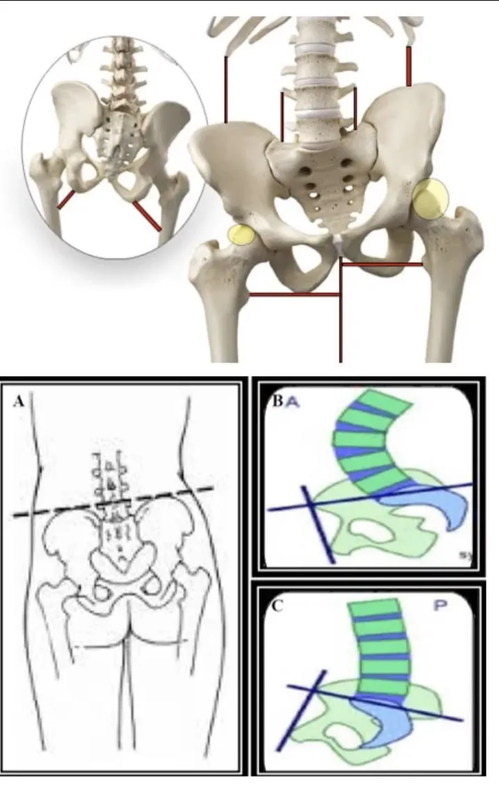
2. Anterior pelvic tilt
The Superior pubic ramusThe upper bony arm of the pubic bone at the front of the pelvis.General meaning, not from the slides lies inferior to the Posterior inferior iliac spineA bony bump at the back of the hip bone (ilium), just below the PSIS.General meaning, not from the slides. Causes:
- Tight Low back extensorsThe muscles along the lower spine that straighten the back and arch it backward.General meaning, not from the slides
- Tight Hip flexorsThe muscles at the front of the hip that lift the thigh forward and up.General meaning, not from the slides
- Lengthened Hip extensorsThe muscles at the back of the hip that move the thigh backward.General meaning, not from the slides
- Excessive LordosisThe normal inward curve of the lower back. Excessive lordosis means the curve is exaggerated.General meaning, not from the slides
3. Posterior pelvic tilt
The superior pubic ramus is superior to the posterior inferior iliac spines. Causes:
- Lumbar VertebraOne of the bones that stack to form the spine.General meaning, not from the slides flexed excessively
- Tight hamstrings
- Elongated low back extensors
Anterior and posterior tilt are mirror images: what is tight in one is lengthened in the other (back extensors, hip flexors ↔ hip extensors/hamstrings).
4. Pelvic rotation
The plumb line falls to the right or left of the gluteal cleft. Causes:
- Tight Medial rotatorsThe muscles that turn the hip or leg inward.General meaning, not from the slides and hip flexors on the rotated side
- Ipsilateral lumbar rotation
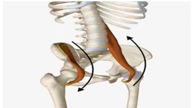
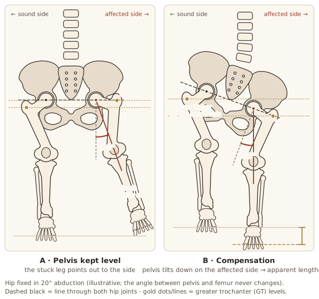
5. Abducted hip
The greater trochanter is higher on the involved side. Causes:
- Tight Hip abductorsThe muscles on the outer hip that lift the leg out to the side.General meaning, not from the slides
- Tight ContralateralOn the opposite side of the body from the structure being discussed.General meaning, not from the slides Hip adductorsThe muscles on the inner thigh that pull the leg toward the midline.General meaning, not from the slides
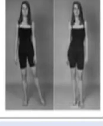
A Fixed abduction deformityA hip held permanently out to the side that cannot be brought back toward the midline.General meaning, not from the slides is CompensatedThe body adjusts another part to make up for a problem elsewhere.General meaning, not from the slides by scoliosis convex toward the affected side and the pelvis tilting down, causing Apparent lengtheningThe limb looks longer, without the bone actually being longer.General meaning, not from the slides of the limb.
Knee Deformities
1. Genu recurvatum
The knee is HyperextensionStraightening a joint beyond its normal limit.General meaning, not from the slides and the Gravity lineThe imaginary vertical line along which body weight is pulled down by gravity.General meaning, not from the slides lies far forward of the joint axis.
Causes:
- Tight QuadricepsThe large muscle group at the front of the thigh that straightens the knee.General meaning, not from the slides, GastrocnemiusThe large calf muscle that points the foot down and helps bend the knee.General meaning, not from the slides and SoleusA flat calf muscle lying under the gastrocnemius that points the foot downward.General meaning, not from the slides
- Stretched PopliteusA small muscle at the back of the knee that helps the knee bend.General meaning, not from the slides and HamstringsThe muscle group at the back of the thigh that bends the knee and extends the hip.General meaning, not from the slides
- Compression forces anteriorly
- Shape of the Tibial plateauThe flat upper surface of the tibia where it meets the femur at the knee.General meaning, not from the slides
Key symptoms of Genu recurvatumA knee that is hyperextended, with the gravity line lying far forward of the joint axis. syndrome:
- Pain in the inner leg or outer back of the knee
- Extension gait pattern
- Poor ProprioceptionThe body's sense of where its joints are and how they are moving.General meaning, not from the slides control of Terminal knee extensionThe last few degrees of straightening the knee.General meaning, not from the slides
- Pinching in front of the knee
- Difficulty with endurance activities
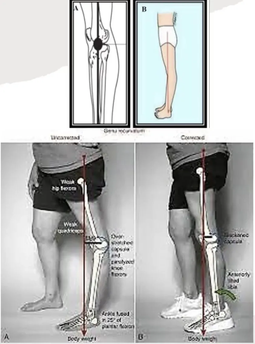
Management
- OrthosisA brace, support or insert worn on the body to protect, align or support a body part.General meaning, not from the slides and bracing — protect and stabilise the knee and limit abnormal hyperextension, allowing active movement and a more harmonious gait.
- Surgery (rare, severe cases): Proximal tibial osteotomyA surgical cut through the upper part of the tibia so that the knee alignment can be changed.General meaning, not from the slides to reduce hyperextension.
- Complications if untreated: continued strain, soft-tissue damage, increasing deformity, and it may lead to Genu valgumKnock knees: the mechanical axis of the lower limb is displaced laterally., Genu varumBow legs: the leg deviates toward the midline relative to the thigh, so the knee lies lateral to the mechanical axis. and knee OsteoarthritisWear-and-tear arthritis in which joint cartilage thins and the joint becomes stiff and painful.General meaning, not from the slides.
- Precautions: avoid activities that strain the knees, follow the PTPhysical therapy (physiotherapy): treatment that uses exercise, movement and hands-on techniques to restore movement and function.General meaning, not from the slides programme strictly, see the doctor regularly.
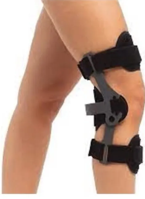
2. Flexed knee
The Plumb lineA string with a weight on the end, hung to give a true vertical reference line for judging posture.General meaning, not from the slides falls posterior to the joint axis.
Causes:
- Hamstring tightness
- Stretched quadriceps
- Tight gastrocnemius
- Posterior compression forces
- Bony and soft-tissue limitations
Clinical presentation
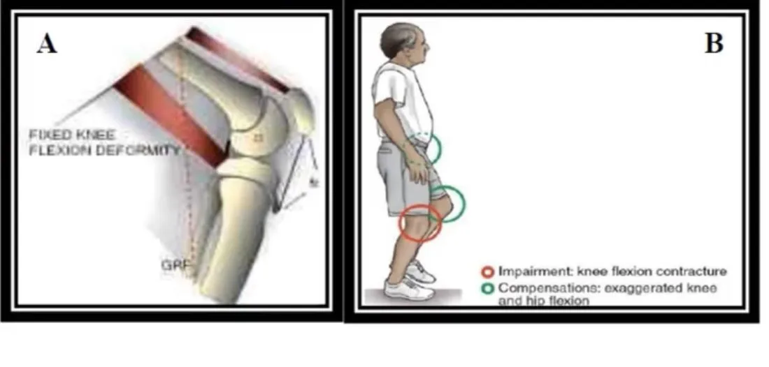
- Flexed kneeA knee held bent, with the plumb line falling behind the joint axis. gait → more strain on the quadriceps and higher PatellofemoralRelating to the kneecap and the femur, and the joint between them.General meaning, not from the slides contact forces.
- Reduced walking distance, quadriceps weakness and early fatigue.
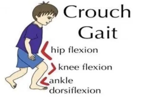
- Anterior knee pain, a progressive Crouch gaitA walking pattern with the hips, knees and ankles all bent (hip flexion, knee flexion and ankle dorsiflexion). (hip flexion + knee flexion + ankle DorsiflexionPulling the foot up toward the shin.General meaning, not from the slides) and limping. A Hip flexion contractureA lasting shortening of the tissues at the front of the hip, so the hip cannot fully straighten.General meaning, not from the slides with LordosisThe normal inward curve of the lower back. Excessive lordosis means the curve is exaggerated.General meaning, not from the slides may follow.
- Early changes: shorter stride, reduced Popliteal angleA measurement of the angle at the back of the knee, used to judge hamstring tightness.General meaning, not from the slides, knee flexed at Initial stanceThe start of the stance phase of walking, when the foot first touches the ground.General meaning, not from the slides and throughout gait.
- Late changes: severe knee and hip ContractureA lasting shortening of muscle or soft tissue that limits how far a joint can move.General meaning, not from the slides and Patella altaA kneecap that sits higher than normal.General meaning, not from the slides.
- Functional problems with Weight bearingPutting body weight through a limb or joint, for example by standing or walking on it.General meaning, not from the slides and bed/chair positioning; daily activities cost more energy.
Physical therapy management
- Manual stretchingStretching done by the therapist's hands.General meaning, not from the slides
- Prolonged stretch through StrappingStraps or tape used to hold a body part in a chosen position.General meaning, not from the slides on a Tilt tableA table that can be tilted from lying toward upright.General meaning, not from the slides
- Sandbag weight over the DistalFarther from the trunk or the point of attachment of a limb.General meaning, not from the slides FemurThe thigh bone, the longest bone in the body.General meaning, not from the slides
- Mechanical tractionA pulling force applied to a joint or limb by a machine or weights.General meaning, not from the slides
- Passive ROMMovement of a joint carried out by someone or something else, with no effort from the patient.General meaning, not from the slides and Joint mobilisationA hands-on technique of gently moving a joint to improve its movement.General meaning, not from the slides
The effect of any treatment depends on the applied TorqueA turning (rotational) force.General meaning, not from the slides and on the duration and frequency of treatment.
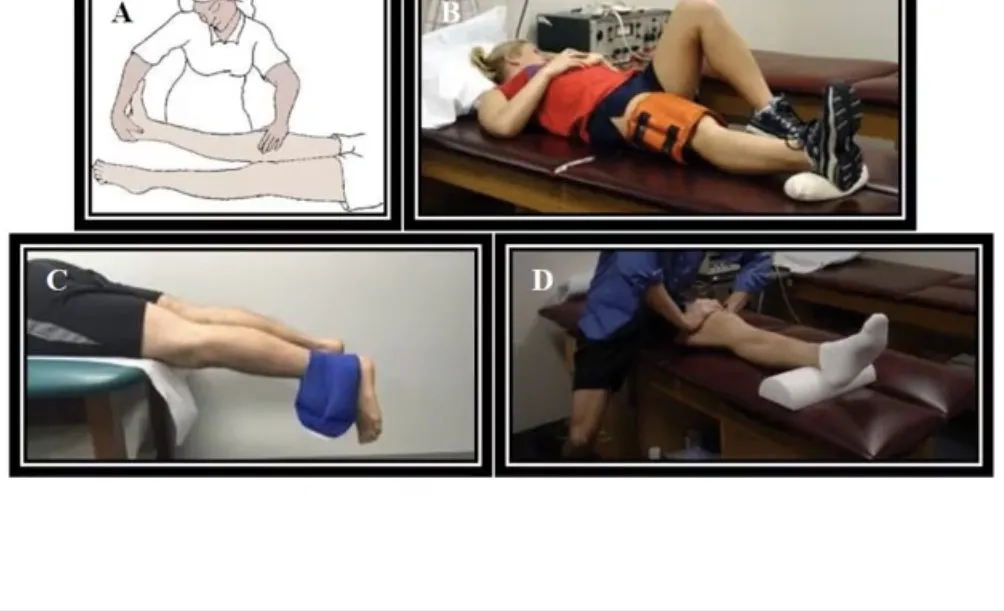
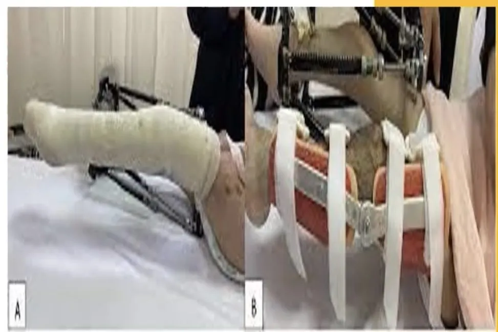
Other methods: orthoses, casting and bracing; splints that apply low stretching force over long periods (some are inflatable, easy to apply and comfortable). In most cases splints and orthoses prevent deformity or maintain Range of motion (ROM)How far a joint can move in each direction.General meaning, not from the slides after stretching — they do not increase ROM.
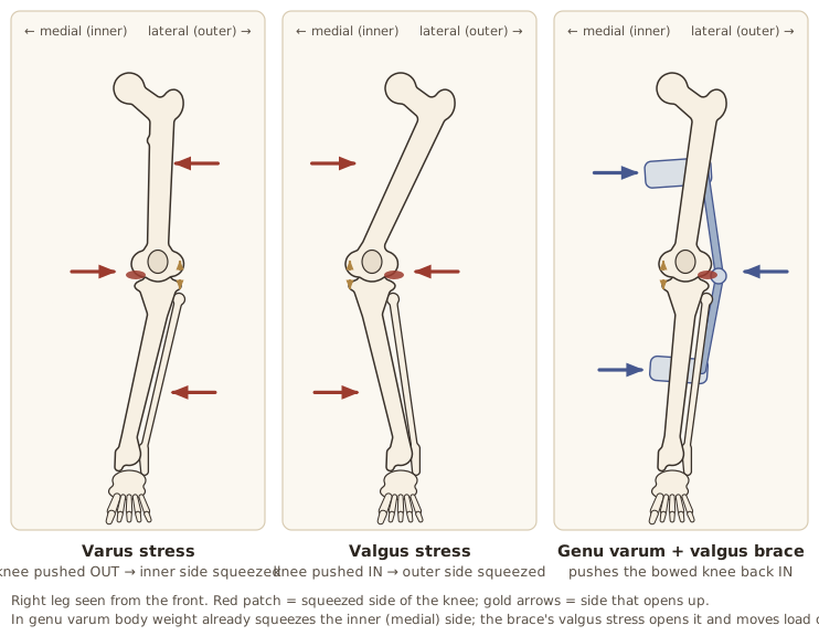
3. Genu varum (bow legs)
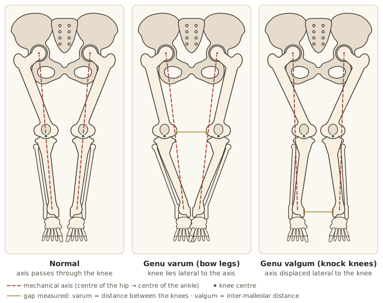
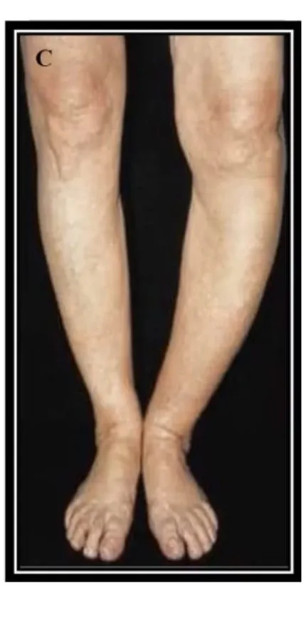
The leg deviates toward the MidlineThe imaginary vertical line that divides the body into left and right halves.General meaning, not from the slides relative to the thigh; the knee joint lies lateral to the Mechanical axisThe imaginary straight line from the centre of the hip to the centre of the ankle, used to judge limb alignment.General meaning, not from the slides of the limb.
Causes:
- Tight Medial rotatorsThe muscles that turn the hip or leg inward.General meaning, not from the slides at the hip
- Compression of medial joint structures
- Femoral retroversionThe femoral head and neck point backward relative to the patellae, so the leg turns outward.
- Elongated Lateral rotatorsThe muscles that turn the hip or leg outward.General meaning, not from the slides and popliteus
Assessment: AngulationThe angle at which a bone or limb bends away from a straight line.General meaning, not from the slides standing/SupineLying face up, on the back.General meaning, not from the slides; distance between the knees.
Surgical options:
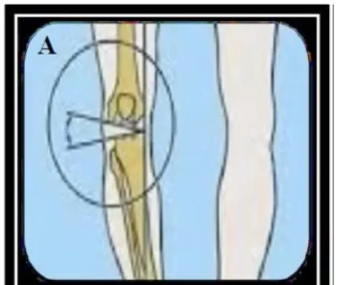
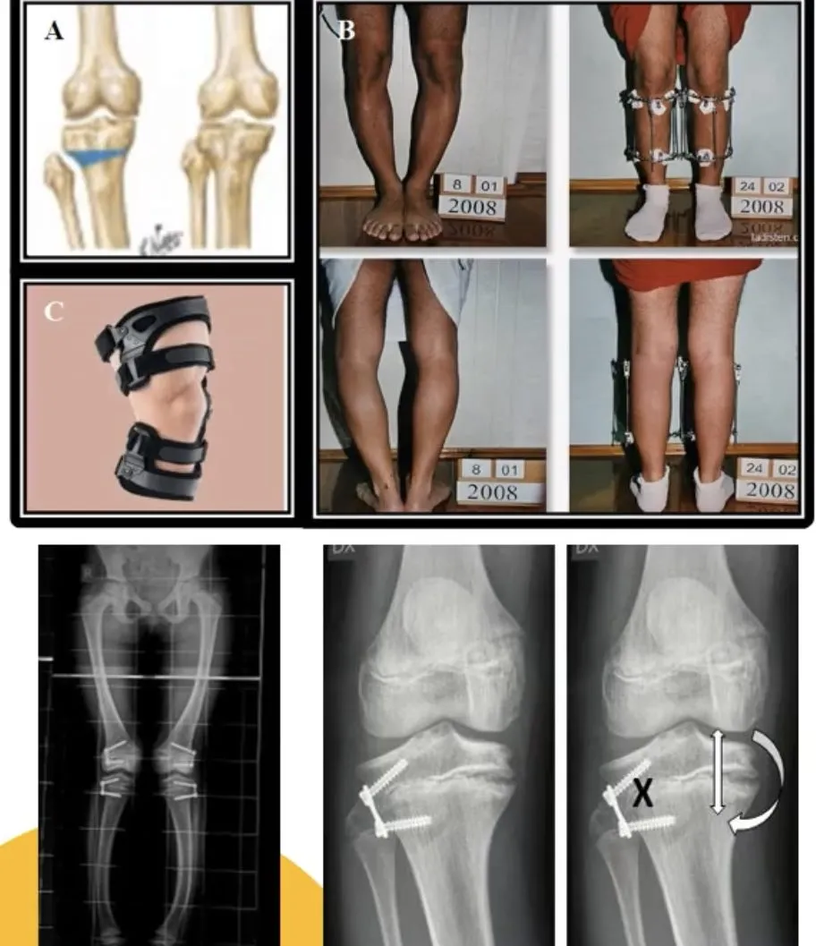
- Epiphyseal staplingA surgical procedure that fixes metal staples across one side of a growth plate to slow its growth there.General meaning, not from the slides at 10–11 years
- Corrective OsteotomyA surgical cut through a bone so that it can be realigned.General meaning, not from the slides at maturity — traditionally a proximal tibial osteotomy
- Ilizarov methodOsteotomy plus an external fixator that allows gradual adjustment (about 24 weeks in genu varum); restores alignment but has a high complication rate. method: osteotomy plus an External fixatorA frame outside the body, attached to a bone by pins or wires, that holds the bone in position.General meaning, not from the slides that allows gradual adjustment over about 24 weeks; restores alignment but with a high complication rate
Bracing: a Compartmental unloaderA knee brace that takes load off one side of the knee. In genu varum it lowers stress on the medial tibiofemoral joint. — a person with genu varum needs a ValgusBent outward: the part beyond a joint angles away from the midline of the body.General meaning, not from the slides orthosis, which lowers stress on the medial Tibiofemoral jointThe main knee joint, between the tibia and the femur.General meaning, not from the slides joint. A compartmental unloader and a Lateral heel wedgeA shoe insert that is thicker along the outer edge of the heel.General meaning, not from the slides both give short-term pain relief and may delay Total knee replacementSurgery that replaces the damaged surfaces of the knee joint with artificial parts.General meaning, not from the slides.
4. Genu valgum (knock knees)
The mechanical axis of the lower limb is displaced laterally.
Causes:
- Tight Iliotibial bandA thick band of connective tissue down the outer thigh, from the hip to just below the knee.General meaning, not from the slides and lateral knee structures
- Femoral anteversionThe forward twist of the femoral neck relative to the knee. Normally 10–15° in adults and about 30–35° in infants.
- Lengthened medial knee structures
- Compression of the lateral knee joint
- Foot PronationIn the foot: rolling inward so the arch flattens. In the forearm: turning the palm downward.General meaning, not from the slides
Assessment: angulation standing/supine; Inter-malleolar distanceThe gap between the inner ankle bones, used to measure knock knees.General meaning, not from the slides.
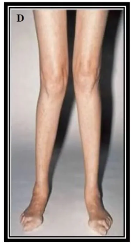
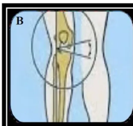
Investigations: X-ray when severe or possibly PathologicalCaused by disease or abnormal change, rather than being part of normal development.General meaning, not from the slides — a standing APAnteroposterior: an X-ray view taken from front to back.General meaning, not from the slides long film (hips to ankles) with PatellaThe kneecap.General meaning, not from the slides facing forward. Look for RicketsA childhood condition in which bones become soft and weak, usually from a lack of vitamin D.General meaning, not from the slides, Tibia vara (Blount's)A growth disorder of the upper tibia that causes the lower leg to bow inward.General meaning, not from the slides (Blount’s) or EpiphysisThe end part of a long bone, separated from the shaft by the growth plate in a child.General meaning, not from the slides injury.
Clinical notes
- Often collapsed Medial longitudinal archThe arch along the inner side of the foot. The normal arch acts as a shock absorber., with the inner ankle bones lower than the outer.
- Uncorrected adult genu valgum → injury and chronic problems such as ChondromalaciaSoftening and wearing of joint cartilage, often under the kneecap.General meaning, not from the slides and osteoarthritis.
- Normal in children aged 2–5 years; almost all resolve with growth.
- Prolonged, pronounced or hereditary cases: orthotic shoes or night leg braces. Persistent/worsening cases: surgery (lower femur adjustment, TKR).
- Weight loss and swapping high-impact for low-impact exercise slow progression — each step loads the knee toward valgus, more so with greater angle or weight.
- Physical therapy is generally of benefit.
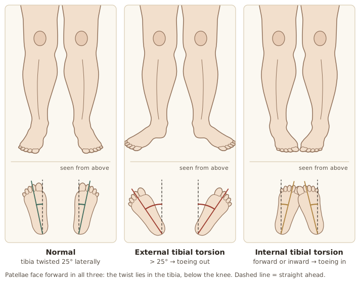
5. External (lateral) tibial torsion
Normally the distal TibiaThe shin bone, the larger bone of the lower leg.General meaning, not from the slides is rotated laterally 25° from the ProximalCloser to the trunk or the point of attachment of a limb.General meaning, not from the slides end. More than 25° is External tibial torsionThe distal tibia is rotated laterally by more than the normal 25° from the proximal end, so the foot toes out. (Out-toeingWalking with the feet turned outward.General meaning, not from the slides).
Causes:
- Tight Tensor fasciae lataeA small muscle at the front and outside of the hip that tightens the iliotibial band.General meaning, not from the slides / iliotibial band
- Bony MalalignmentBones or joints that are not lined up in their normal position.General meaning, not from the slides
- Cruciate ligamentOne of the two crossing ligaments inside the knee that keep the tibia and femur stable.General meaning, not from the slides tear
- Femoral retroversion
6. Internal tibial torsion
The feet face directly forward or inward (In-toeingWalking with the feet turned inward.General meaning, not from the slides).
Causes:
- Tight Medial hamstringsThe hamstring muscles on the inner side of the back of the thigh.General meaning, not from the slides and GracilisA thin muscle on the inner thigh.General meaning, not from the slides
- Structural tibial deformity (traumatic or developmental)
- ACLAnterior cruciate ligament: the knee ligament that stops the tibia sliding forward on the femur.General meaning, not from the slides tear
- Femoral anteversion
- Foot pronation
- Genu valgus
Management of Rotational Deformities (In-toeing)
| Approach | Verdict |
|---|---|
| Wait / observe | Best — corrects spontaneously in the vast majority as the limb External rotationTurning a limb outward, away from the midline of the body.General meaning, not from the slides with growth; convince the family that observation is appropriate |
| Shoe wedges and inserts | Ineffective |
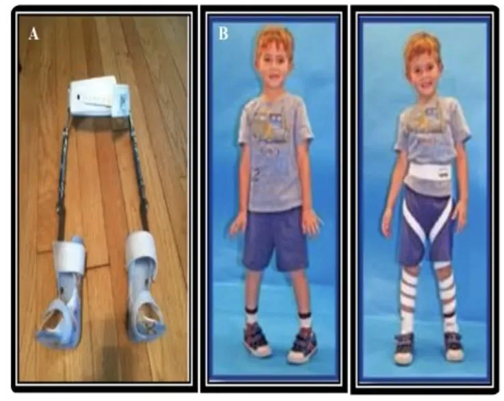
| Bracing with Twister cablesCables attached to the shoes and a band around the pelvis that apply a gentle twisting force to the legs.General meaning, not from the slides | Ineffective and limits activity |
| Night splintA splint worn only during sleep.General meaning, not from the slides | Better tolerated — benefit uncertain |
- Only < 1% of femoral and TibiaThe shin bone, the larger bone of the lower leg.General meaning, not from the slides TorsionA twist of a bone along its length.General meaning, not from the slides deformities fail to resolve and may need surgery in late childhood.
- Twister cables and De-rotation strapsElastic bands attached to the shoe or AFO that wind up the leg to a mesh pelvic band. are used for rotational issues, mainly in children. De-rotation straps are elastic bands attached to the shoe or AFOAnkle-foot orthosis: a brace that runs from the lower leg to the foot to support the ankle and foot.General meaning, not from the slides that wind up the leg to a mesh pelvic band.
Surgery is indicated in an older child (> 8–10 years) with significant functional disability.
Ankle and Foot
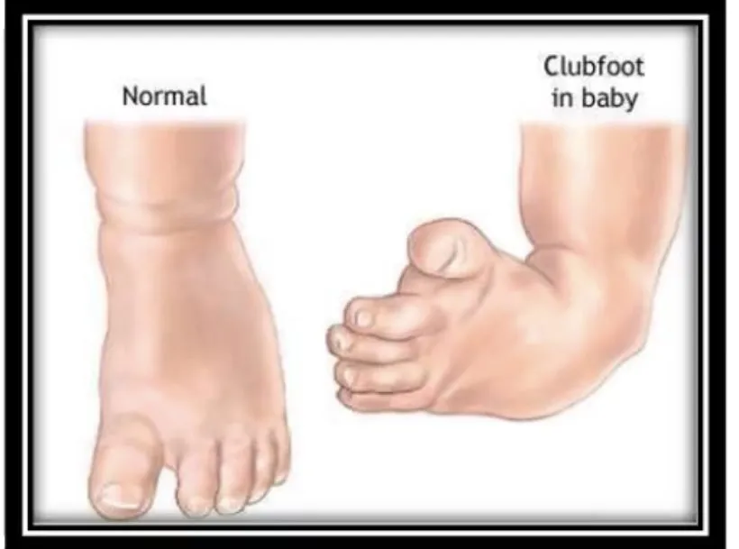
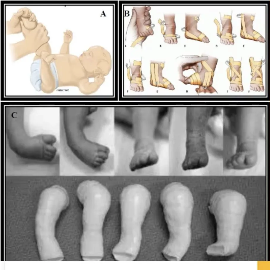
I. Club foot — Talipes Equino-Varus (TEV)
- One of the commonest foot deformities; involves the ankle, Subtalar jointThe joint just below the ankle, between the talus and the heel bone.General meaning, not from the slides and Midtarsal jointThe joints in the middle of the foot, between the hindfoot and the forefoot.General meaning, not from the slides joints.
- A 3-dimensional deformity: Plantar flexionPointing the foot downward, away from the shin. (EquinusThe foot is pointed downward (plantar flexed), like a horse's hoof.General meaning, not from the slides), InversionTurning the sole of the foot inward.General meaning, not from the slides (VarusBent inward: the part beyond a joint angles toward the midline of the body.General meaning, not from the slides) and AdductionMoving a limb toward the midline of the body.General meaning, not from the slides.
- Clinically the sole faces inward, giving an L-shaped lower leg.
- IncidenceHow often a condition occurs in a population.General meaning, not from the slides: 1–2 per 1000 live births; males twice as affected; 50% BilateralAffecting both sides of the body.General meaning, not from the slides.
Types
| Structural (fixed) | Postural (mobile) |
|---|---|
| Usually treated surgically | Usually treated Conservative treatmentTreatment without surgery, such as exercise, stretching, bracing or casting.General meaning, not from the slides |
| Cannot be corrected passively | Can be corrected passively |
| Genetic; runs in families | Usually with another birth condition such as Spina bifidaA birth defect in which part of the spine does not close fully.General meaning, not from the slides |
Conservative management
- Rhythmic, repeated gentle ManipulationHands-on movement of a joint or body part by the therapist.General meaning, not from the slides
- StrappingStraps or tape used to hold a body part in a chosen position.General meaning, not from the slides
- POP (plaster of Paris)Plaster of Paris: a white powder that sets hard when wet, used to make casts.General meaning, not from the slides (plaster of Paris) cast — Serial castingA series of casts applied one after another, each changing the position a little to correct a deformity gradually.General meaning, not from the slides
- Education and instructions to the parents
Instructions to parents: comply with treatment; understand correction is gradual; watch the baby frequently for unusual crying, swelling and discoloration; keep the cast/strapping away from fluids, urine and stool.
Precautions when applying cast or strapping: moderate pressure to keep circulation; enough cotton around the MalleolusThe bony bump on each side of the ankle.General meaning, not from the slides to avoid Pressure soreDamage to the skin and tissue under it caused by prolonged pressure.General meaning, not from the slides; keep the limb clean; avoid skin wrinkles.
II. Pes planus (pronated / flat foot)
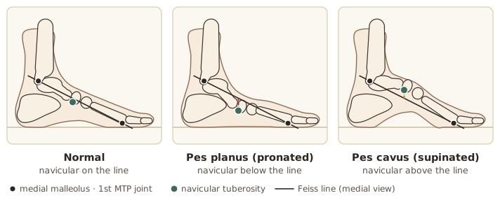
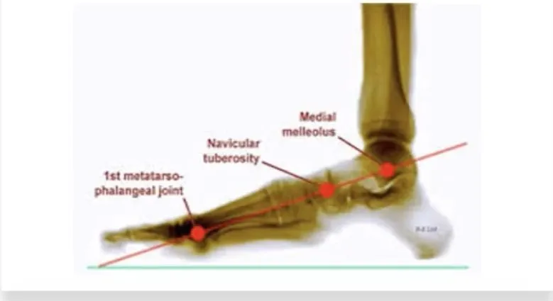
- Decreased Medial longitudinal archThe arch along the inner side of the foot. The normal arch acts as a shock absorber., Achilles tendonThe thick tendon at the back of the ankle that joins the calf muscles to the heel bone.General meaning, not from the slides ConvexityThe outward-bulging side of a curve.General meaning, not from the slides medially, and the Navicular tuberosityThe bony bump of the navicular on the inner side of the foot.General meaning, not from the slides lies below the Feiss lineA line from the medial malleolus to the first MTP joint. The navicular tuberosity lies below it in flat foot and above it in pes cavus. (line from medial malleolus to 1st MTP jointMetatarsophalangeal joint: the joint at the base of a toe, where the toe meets the foot bone.General meaning, not from the slides joint).
- Causes: shortened Peroneal musclesThe muscles on the outer side of the lower leg that turn the foot outward.General meaning, not from the slides muscles; elongated Tibialis posteriorA deep calf muscle whose tendon runs behind the inner ankle bone and helps support the foot arch.General meaning, not from the slides; stretched spring (Spring ligamentPlantar calcaneonavicular ligament: a ligament under the foot between the heel bone and the navicular that supports the arch.General meaning, not from the slides) ligament; structural displacement of TalusThe ankle bone that sits between the tibia above and the heel bone below.General meaning, not from the slides, CalcaneusThe heel bone.General meaning, not from the slides and NavicularA boat-shaped bone on the inner side of the midfoot.General meaning, not from the slides.
- Normal development: Pes planusFlat foot: decreased medial longitudinal arch, with the navicular tuberosity lying below the Feiss line. are normal when learning to walk; the medial arch does not develop until the 2nd–3rd year; the normal arch acts as a shock absorber.
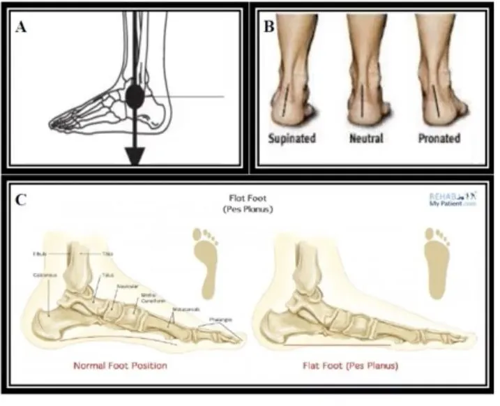
Investigation: ask the patient to stand on tiptoe — if the arch restores itself on tiptoe or when unloaded, no action is needed (flexible flat foot).
Contributing factors: improper footwear (high heels, shoes limiting toe movement), obesity, tight Achilles tendon, Ligamentous laxityLooseness of the ligaments, so joints move more than normal.General meaning, not from the slides.
Diagnosis (standing): loss of medial arch, Heel valgusThe heel bone tilts outward, away from the midline.General meaning, not from the slides, SupinationIn the foot: rolling outward so the arch rises. In the forearm: turning the palm upward.General meaning, not from the slides ForefootThe front part of the foot: the toes and the long bones behind them.General meaning, not from the slides. Standing foot X-ray: flat arch, heel valgus.
Treatment
- No treatment if symmetrical, bilateral, painless and not progressing.
- Non-surgical: heel-cord stretching; OrthosisA brace, support or insert worn on the body to protect, align or support a body part.General meaning, not from the slides (Heel wedgeA shoe insert that is thicker on one side of the heel, used to tilt the heel.General meaning, not from the slides or insoles) to correct calcaneal valgus or support the arch; reduce contributing factors (lose weight, proper footwear).
- Surgical indications: Cerebral palsyA group of lifelong movement and posture disorders caused by damage to the developing brain.General meaning, not from the slides, fixed and painful pes planus, Posterior tibial tendon dysfunctionThe posterior tibial tendon stretches out and becomes incompetent, so that a long-standing flatfoot becomes symptomatic (acquired adult flatfoot deformity)., failed conservative treatment.
- Procedures: Achilles tendon lengtheningSurgery that lengthens the Achilles tendon.General meaning, not from the slides, Calcaneal osteotomyA surgical cut through the heel bone so that it can be realigned.General meaning, not from the slides, Tibialis posterior tendon reconstructionSurgery that repairs or rebuilds the tibialis posterior tendon.General meaning, not from the slides, Hindfoot fusionSurgery that joins the bones of the hindfoot together so they no longer move separately.General meaning, not from the slides for pain relief, Subtalar arthrodesisSurgery that fuses the subtalar joint so its bones become one.General meaning, not from the slides to correct deformity.
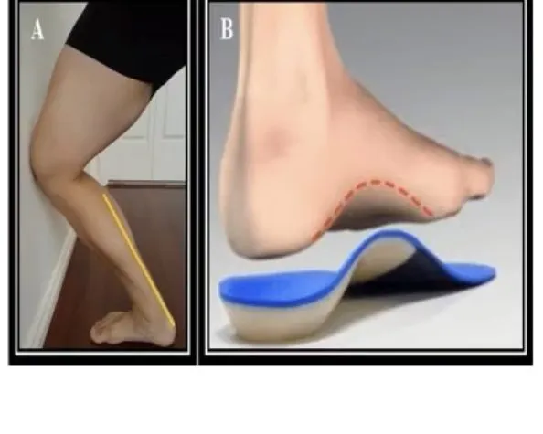
Acquired Adult Flatfoot Deformity (Posterior Tibial Tendon Dysfunction)
A symptomatic flatfoot:
- Pain mainly on the medial ankle, sometimes lateral HindfootThe back part of the foot: the heel bone and the ankle bone above it.General meaning, not from the slides from bony ImpingementPinching or jamming of bone or soft tissue against another structure.General meaning, not from the slides.
- Becomes symptomatic after years of flatfoot, when medial structures including the posterior tibial tendon stretch out and become IncompetentNot working properly; here, a stretched tendon that can no longer do its job.General meaning, not from the slides.
- Stage 2: the patient cannot do a Single-leg heel raiseStanding on one leg and rising up onto the ball of the foot.General meaning, not from the slides (which needs a functional posterior tibial tendon). The tendon is stretched beyond working length but not ruptured.
Protocol — two components:
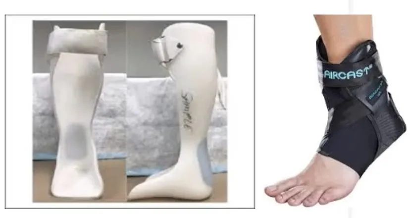
- Bracing to protect the injured medial structures.
- A high-repetition, low-resistance strengthening programme for the posterior tibial tendon.
Limitations: it does not fix the deformity or undo tendon damage, but improves symptoms and function. Patients risk recurrence and will likely need a scaled-down routine 3–4×/week for life; high-demand activities such as running may be hard.
Bracing component: Stage 2 → a custom-moulded AFOAnkle-foot orthosis: a brace that runs from the lower leg to the foot to support the ankle and foot.General meaning, not from the slides. Some find it too hot, so the Aircast posterior tibial ankle lacerAn over-the-counter lace-up ankle brace, usable immediately, offered as an alternative to the custom-moulded AFO. (over the counter, usable immediately) is an alternative. After a few months, move to an over-the-counter orthotic with medial arch support. Wear the brace/orthotic whenever walking.
Exercise component:
| Phase | Start criterion | Content | Duration |
|---|---|---|---|
| 1 | — | Sole-to-sole exerciseSitting with the feet dangling and turning the soles inward to bring them together, using gravity only. exercises (sit, feet dangling, invert to bring soles together — gravity only) or picking up a ball with the heels; start 4 × 10, increase daily to 12 × 25 | ~10–14 days |
| 2 | Comfortably 12 × 25 of a Phase 1 exercise | Phase 1 as warm-up; Resistance bandAn elastic band that you pull or stretch against to add resistance to an exercise.General meaning, not from the slides inversion/EversionTurning the sole of the foot outward.General meaning, not from the slides/DorsiflexionPulling the foot up toward the shin.General meaning, not from the slides (inversion most important), controlled EccentricA muscle contraction in which the muscle lengthens while it works, such as controlled lowering.General meaning, not from the slides return, no leg rotation, 4 × 10 → 10 × 20 (200 total); Double-leg heel riseStanding on both legs and rising up onto the balls of the feet.General meaning, not from the slides with the uninvolved leg taking 75–85% of body weight, 4 × 5 → 10 × 20 | 4–8 weeks; back off a few days if symptoms flare |
| 3 | Comfortably 200 reps of band work and the heel rises | Phase 1 warm-up; stiffer band, 200–300 reps; double-leg heel rises with the uninvolved leg taking only 50%; single-leg heel rises; toe walking 10 ft, knees straight, progressing to 5 × 100 ft | 4–8 weeks |
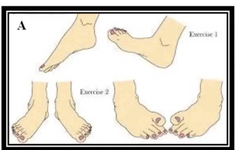
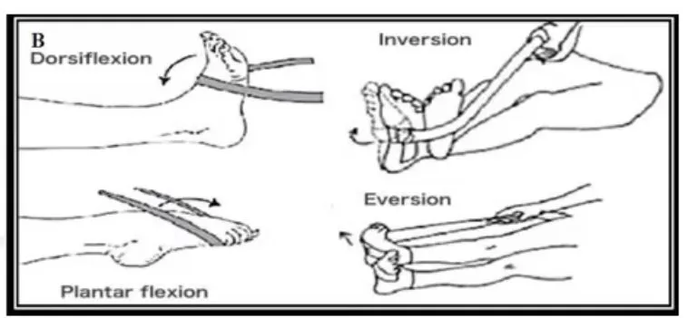
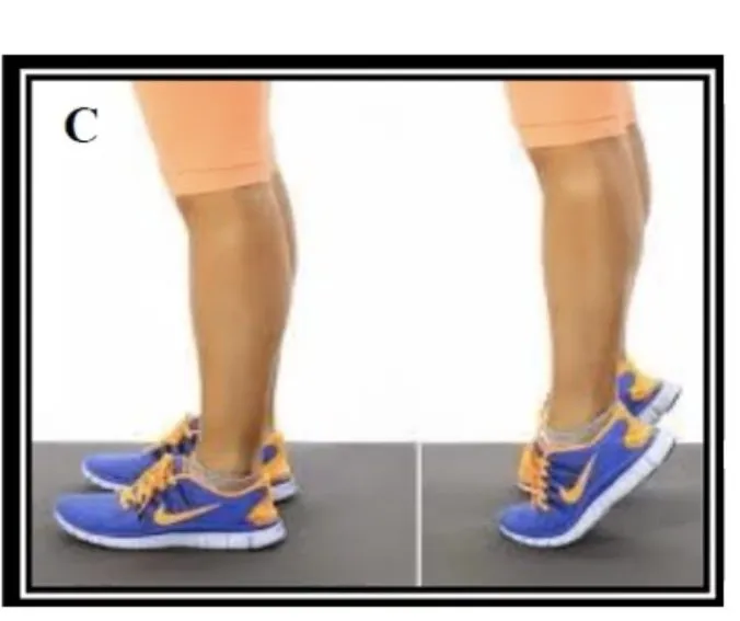
Stabilisation: afterwards, a 10–15 minute routine of double-leg heel rises, band work and toe walking, performed a minimum of 3 times.
III. Pes cavus (supinated / high arch)
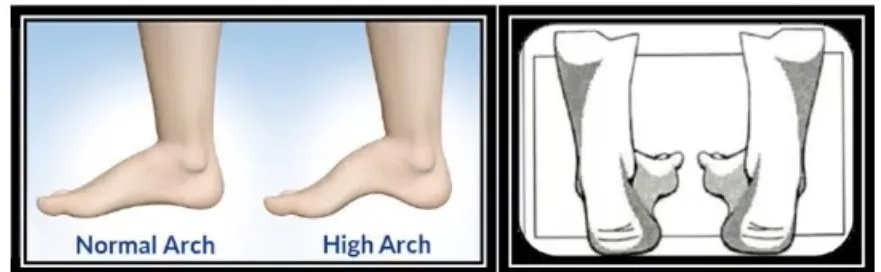
- High medial longitudinal arch; navicular lies above Feiss’ line.
- Causes: shortened posterior and Anterior tibial musclesThe muscle at the front of the shin (tibialis anterior) that lifts the foot upward.General meaning, not from the slides muscles; elongated peroneals and lateral ligaments.
Pes cavusHigh-arched foot: a high medial longitudinal arch, with the navicular lying above the Feiss line. is the mirror image of pes planus: tibialis posterior is shortened in cavus but elongated in planus; the peroneals are elongated in cavus but shortened in planus.
IV. Hallux valgus
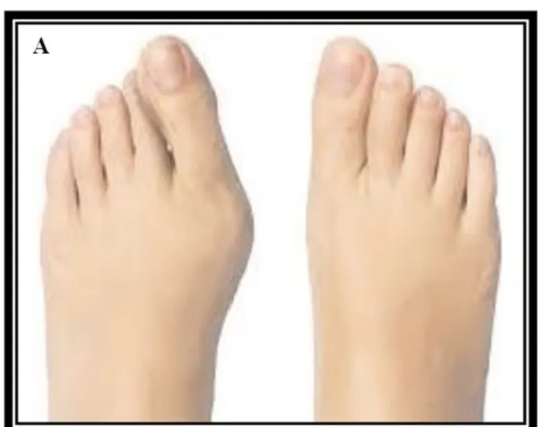
Lateral deviation of the great toe at the metatarsophalangeal joint. Causes:
- Excessive medial bone growth of the first MetatarsalOne of the five long bones in the middle of the foot, between the ankle bones and the toes.General meaning, not from the slides head
- Joint DislocationA joint whose bones have been forced out of their normal alignment.General meaning, not from the slides
- Tight Adductor hallucisA small foot muscle that pulls the big toe toward the other toes.General meaning, not from the slides
- Stretched Abductor hallucisA small foot muscle that pulls the big toe away from the other toes.General meaning, not from the slides
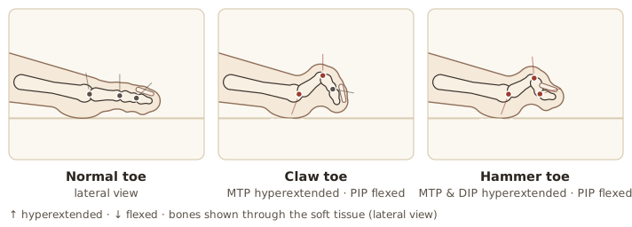
V. Claw toes
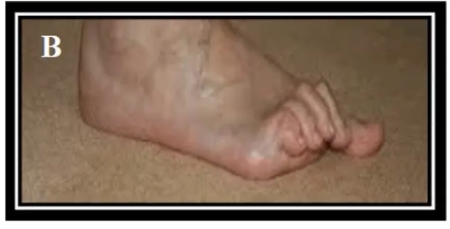
MTP HyperextensionStraightening a joint beyond its normal limit.General meaning, not from the slides + PIP jointProximal interphalangeal joint: the middle joint of a finger or toe.General meaning, not from the slides flexion, associated with pes cavus. Causes: tight Long toe flexorsThe muscles that curl the toes downward.General meaning, not from the slides; short Toe extensorsThe muscles that straighten the toes and lift them upward.General meaning, not from the slides.
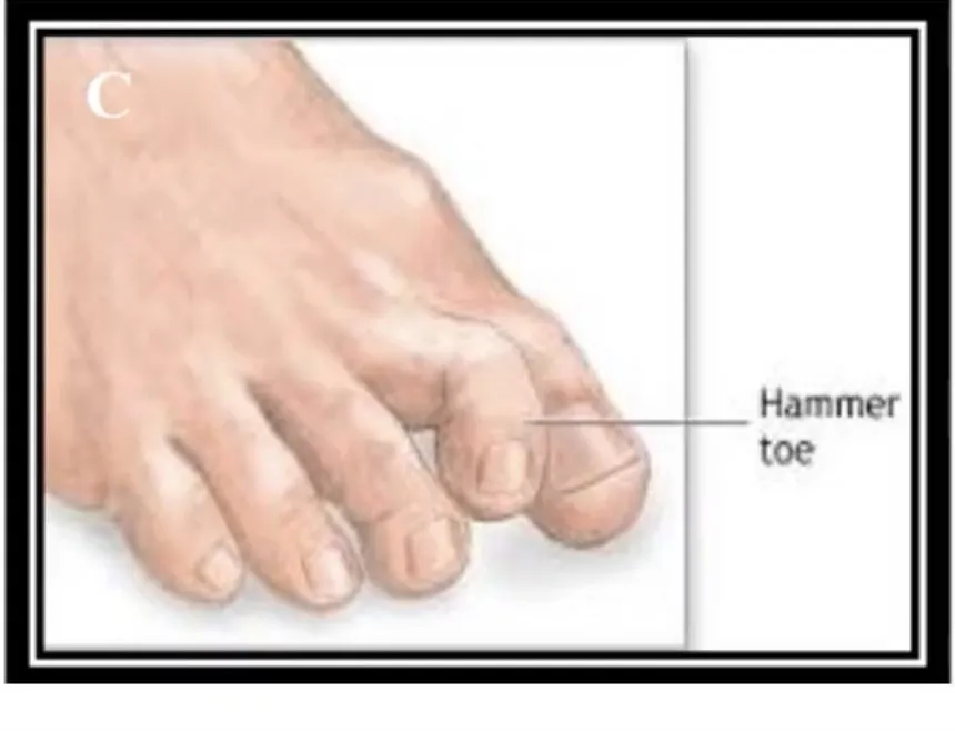
VI. Hammer toes
MTP and DIP jointDistal interphalangeal joint: the joint nearest the tip of a finger or toe.General meaning, not from the slides hyperextension + PIP flexion. Causes: short toe extensors; lengthened LumbricalsSmall muscles of the foot (and hand) that help bend the base joint of each toe and straighten the end joints.General meaning, not from the slides.
| Toe deformity | MTP | PIP | DIP |
|---|---|---|---|
| Claw toesMTP hyperextension with PIP flexion, associated with pes cavus. | Hyperextended | Flexed | — |
| Hammer toesMTP and DIP hyperextension with PIP flexion. | Hyperextended | Flexed | Hyperextended |
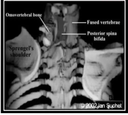
Scapular Deformity — Congenital Elevation of the Scapula (Sprengel’s shoulder)
- Due to arrest of the Caudal migrationThe downward movement (toward the tail end of the body) of the shoulder blade during development.General meaning, not from the slides of the ScapulaThe shoulder blade.General meaning, not from the slides during development.
- Functional and cosmetic impairment proportional to how high the scapula sits; may include an Omovertebral boneAn abnormal extra bone linking the shoulder blade to the spine.General meaning, not from the slides, fused VertebraOne of the bones that stack to form the spine.General meaning, not from the slides and posterior Spina bifidaA birth defect in which part of the spine does not close fully.General meaning, not from the slides.
- Muscle defects: TrapeziusA large, flat, triangular muscle across the upper back and neck that moves and steadies the shoulder blade.General meaning, not from the slides absent or weak; RhomboidsMuscles between the shoulder blade and the spine that pull the shoulder blade toward the spine.General meaning, not from the slides and Levator scapulaeA muscle at the side of the neck that lifts the shoulder blade.General meaning, not from the slides FibrosedTurned into fibrous, scar-like tissue.General meaning, not from the slides and HypoplasticUnderdeveloped.General meaning, not from the slides.
- Aims: correct the deformity (CosmesisImprovement of appearance.General meaning, not from the slides) and improve function (shoulder AbductionMoving a limb away from the midline of the body.General meaning, not from the slides).
- Options: observation for mild cases with minor dysfunction; surgery for severe cases with significant cosmetic and functional problems.
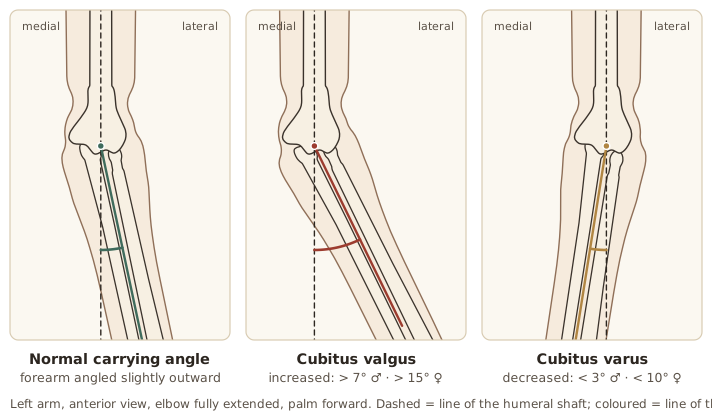
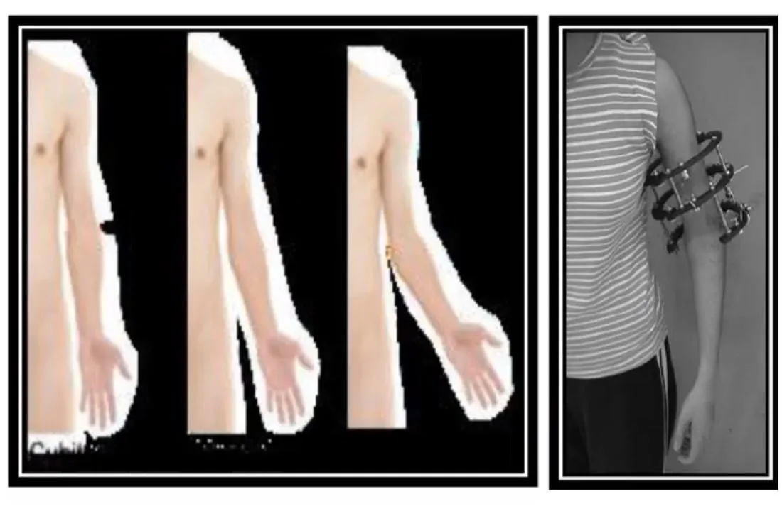
Elbow Deformities — Cubitus Valgus and Cubitus Varus
| Definition (HumerusThe upper-arm bone.General meaning, not from the slides–UlnaThe forearm bone on the little-finger side.General meaning, not from the slides shaft angle) | |
|---|---|
| Cubitus valgusIncreased humerus–ulna shaft angle at the elbow: more than 7° in males and 15° in females. | Increased angle: > 7° in males, > 15° in females |
| Cubitus varusDecreased humerus–ulna shaft angle at the elbow: less than 3° in males and 10° in females. | Decreased angle: < 3° in males, < 10° in females |
- Most obvious with the arm in full extension.
- The humerus–ulna angle determines the type.
- Rule out sinister causes such as Supracondylar fractureA break of the lower end of the humerus, just above the elbow joint.General meaning, not from the slides.
Finger Deformities
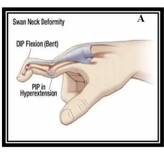
I. Swan-neck deformity
PIP jointProximal interphalangeal joint: the middle joint of a finger or toe.General meaning, not from the slides HyperextensionStraightening a joint beyond its normal limit.General meaning, not from the slides with DIP jointDistal interphalangeal joint: the joint nearest the tip of a finger or toe.General meaning, not from the slides flexion. True Swan-neck deformityA finger deformity with PIP hyperextension and DIP flexion. does not occur in the thumb.
- Commonest cause: Rheumatoid arthritisA long-term disease in which the immune system inflames the joints.General meaning, not from the slides — about 50% of people with RA develop it (RA also causes Ulnar deviationThe fingers drift sideways toward the little-finger (ulnar) side.General meaning, not from the slides, Boutonnière deformityA finger deformity from injury to the tendon that straightens the PIP: the PIP will not straighten while the fingertip (DIP) bends back., Z-thumbA thumb deformity in which the thumb bends into a zig-zag, Z shape.General meaning, not from the slides, and Hammer toesMTP and DIP hyperextension with PIP flexion.).
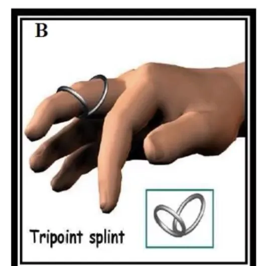
- Non-surgical treatment works best when the PIP is supple:
- PTPhysical therapy (physiotherapy): treatment that uses exercise, movement and hands-on techniques to restore movement and function.General meaning, not from the slides/OTOccupational therapy: treatment that helps people regain the skills for daily activities.General meaning, not from the slides — stretching, massage, strengthening and Joint mobilisationA hands-on technique of gently moving a joint to improve its movement.General meaning, not from the slides.
- A splint (e.g. Tripoint (ring) splintA splint that keeps the PIP joint aligned, blocks hyperextension and still lets the joint bend. / ring splint) that keeps the PIP aligned, blocks hyperextension and still lets it bend.
II. Boutonnière deformity
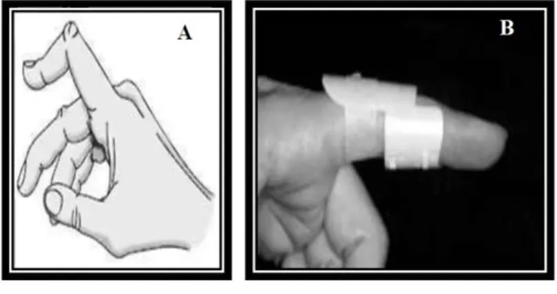
Results from injury to the tendon that straightens the PIP: the PIP will not straighten while the fingertip (DIP) bends back. Untreated, it can become permanent.
Non-surgical treatment:
- Splint at the middle joint to straighten it.
- Exercises — stretching for strength and flexibility.
- Protection — protective splinting or taping for several weeks in sport.
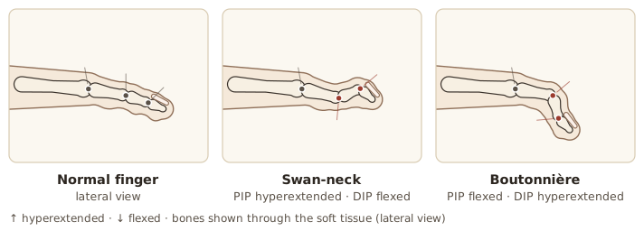
| Finger deformity | PIP | DIP |
|---|---|---|
| Swan neck | Hyperextended | Flexed |
| Boutonnière | Flexed | Hyperextended |