Meniscus Injury: Anatomy, Biomechanics, Assessment, Management and Rehabilitation
Quiz this lecture (126 questions)Prof. Dr Akram Fathy’s lecture covers the MeniscusOne of two semicircular fibrocartilaginous structures between the femoral condyles and the tibial plateau of the knee: the medial and the lateral meniscus. of the knee in six parts: (1) anatomy and blood supply, (2) BiomechanicsHow forces act on and through the body during movement.General meaning, not from the slides (seven functions), (3) mechanism of injury, (4) clinical assessment, (5) classification and management, and (6) RehabilitationExercise and treatment that restore function after injury or surgery.General meaning, not from the slides and Return to sport (RTS)Going back safely to training and competition, judged using return-to-sport criteria..
Objectives and Roadmap
Lecture aim
To equip students with the knowledge and clinical reasoning needed to assess, diagnose and manage MeniscusOne of two semicircular fibrocartilaginous structures between the femoral condyles and the tibial plateau of the knee: the medial and the lateral meniscus. injuries in athletes and active patients, from anatomy and BiomechanicsHow forces act on and through the body during movement.General meaning, not from the slides through evidence-based treatment, RehabilitationExercise and treatment that restore function after injury or surgery.General meaning, not from the slides and safe Return to sport (RTS)Going back safely to training and competition, judged using return-to-sport criteria..
Objectives
By the end of the lecture, students should be able to:
- Describe the anatomy and structural characteristics of the medial and lateral menisci of the knee.
- Explain the functions and biomechanical roles of the menisci in normal knee joint function.
- Discuss the common mechanisms, risk factors, and classifications of meniscal injuries.
- Perform a comprehensive clinical assessment of patients with suspected meniscal tears using history-taking, Physical examinationThe hands-on examination of the patient by the clinician.General meaning, not from the slides, and diagnostic tests.
- Develop evidence-based treatment and rehabilitation plans, considering both Conservative managementTreatment without surgery.General meaning, not from the slides and surgical management.
- Apply evidence-based return-to-sport criteria to determine an athlete’s readiness for safe return to training and competition.
Roadmap
| Part | Topic | Covers | Objective |
|---|---|---|---|
| 01 | Anatomy and blood supply | Structure, attachments, medial vs lateral, VascularHaving a blood supply.General meaning, not from the slides zones | 1 |
| 02 | Biomechanics | Seven functions that protect the Articular cartilageThe smooth cartilage that covers the ends of the bones inside a joint.General meaning, not from the slides | 2 |
| 03 | Mechanism of injury | EpidemiologyThe study of how common a condition is and who it affects.General meaning, not from the slides, TraumaticCaused by a sudden injury.General meaning, not from the slides vs DegenerativeCaused by gradual wear and breakdown of tissue over time.General meaning, not from the slides tears | 3 |
| 04 | Clinical assessment | Presentation, examination, special tests, MRIMagnetic resonance imaging: a scan that shows soft tissues such as menisci and ligaments.General meaning, not from the slides grading | 4 |
| 05 | Classification and management | Tear patterns, preservation, conservative vs surgical care | 3 and 5 |
| 06 | Rehabilitation and return to sport | Three pathways, Weight bearingPutting body weight through the leg.General meaning, not from the slides, strengthening, RTS criteria | 5 and 6 |
Anatomy of the Menisci
Anatomical background
The MeniscusOne of two semicircular fibrocartilaginous structures between the femoral condyles and the tibial plateau of the knee: the medial and the lateral meniscus. are two SemilunarShaped like a half-moon or crescent.General meaning, not from the slides FibrocartilageTough, springy cartilage reinforced with collagen fibers, able to cope with both squeezing and pulling forces.General meaning, not from the slides structures located between the Femoral condylesThe two rounded knobs at the lower end of the femur that sit on top of the tibia and menisci.General meaning, not from the slides and the Tibial plateauThe flat top surface of the tibia on which the femur and the menisci rest.General meaning, not from the slides. They are firmly attached to the TibiaThe shin bone, the larger bone of the lower leg.General meaning, not from the slides through the anterior and posterior Meniscal rootThe anterior or posterior end where the meniscus is firmly anchored to the tibial plateau. Root attachments fix each horn to the tibia. and connected to the Joint capsuleThe fibrous sleeve that surrounds a joint and holds it together.General meaning, not from the slides by the Coronary (meniscotibial) ligamentsLigaments that connect the periphery of the meniscus to the joint capsule..
Essential for normal knee function by:
- Increasing joint CongruencyHow well two joint surfaces fit together. A better fit gives a more stable joint. and stability
- Distributing loads across the Tibiofemoral jointThe main knee joint, where the femur meets the tibia.General meaning, not from the slides
- Absorbing Shock absorptionSoaking up and spreading out impact forces from walking, running and jumping.
- Reducing Contact stressThe force spread over the touching surfaces of femur and tibia. A larger contact area means less stress on the cartilage.General meaning, not from the slides on Articular cartilageThe smooth cartilage that covers the ends of the bones inside a joint.General meaning, not from the slides
- Assisting joint LubricationReducing friction between surfaces with a slippery fluid. and nutrition
- Contributing to ProprioceptionThe body's sense of joint position and movement.General meaning, not from the slides
This list has six items, but the roadmap (slide 3), the Part 2 divider (slide 14) and slides 15–18 speak of seven functions (load distribution and Hoop stressCompression pushes the meniscus outward and its circumferential fibers resist by developing tensile force, turning vertical load into circumferential tension. are counted as functions 1 and 2, and Load sharingThe share of the compressive load carried by the menisci, which rises as the knee bends. is function 4). Both numbers are on the slides; the seven-function list is the one in Part 2.

The slide figure is a labelled front view of the knee: PatellaThe kneecap, a small bone at the front of the knee.General meaning, not from the slides (kneecap), FemurThe thigh bone.General meaning, not from the slides, LCLLateral collateral ligament: the ligament on the outer side of the knee.General meaning, not from the slides (LCL), MCLMedial collateral ligament: the ligament on the inner side of the knee. Its deep fibers attach firmly to the medial meniscus.General meaning, not from the slides (MCL), articular cartilage, lateral meniscus, medial meniscus, ACLAnterior cruciate ligament: a knee ligament that stops the tibia sliding forward on the femur.General meaning, not from the slides (ACL), PCLPosterior cruciate ligament: a knee ligament that stops the tibia sliding backward on the femur.General meaning, not from the slides (PCL), FibulaThe thinner bone running beside the tibia on the outer side of the lower leg.General meaning, not from the slides and tibia.
Gross anatomy
Each meniscus has five parts:
- Horn of the meniscusThe front (anterior horn) or back (posterior horn) end of the crescent-shaped meniscus.General meaning, not from the slides
- Body
- Posterior horn
- Anterior root attachment
- Posterior root attachment
Shared structural characteristics:
| Feature | Description |
|---|---|
| Crescent shape | Both menisci are semilunar (crescent) in shape. |
| Triangular profile | Triangular cross-section, thick at the PeripheryThe outer rim of the meniscus, where it attaches to the joint capsule.General meaning, not from the slides and thin at the Free edgeThe thin inner border of the meniscus that faces the middle of the joint.General meaning, not from the slides. |
| Circumferential fibersCollagen fibers running around the curve of the meniscus. They give tensile strength and resist the outward push of load. | Predominantly circumferential collagen fibers provide Tensile forceA pulling force that stretches a material. Tensile strength is how much pulling it can resist.General meaning, not from the slides. |
| Type I collagenThe type of collagen that makes up most of the dense extracellular matrix of the meniscus. matrix | Dense Extracellular matrixThe material surrounding cells that gives a tissue its structure and strength.General meaning, not from the slides composed primarily of Type I collagen. |
Slide 6 says both menisci are crescent shaped, but slide 9 describes the lateral meniscus as nearly circular (O-shaped) and the medial as C-shaped. Both statements are on the slides, so the difference is flagged, not resolved.
Collagen architecture and hoop stress

How hoop stress works:
- Body weight presses the femur against the tibia and CompressionSqueezing together, such as body weight pressing the femur down on the meniscus. In swelling control it also means pressing on the area, for example with a bandage.General meaning, not from the slides the meniscus.
- Compression tends to push the meniscus outward.
- Circumferential fibers stretch and develop tensile force that resists this DisplacedMoved out of its normal position.General meaning, not from the slides: the hoop-stress mechanism.
Radial tie fibersFibers running from the inner toward the outer border, crossing the circumferential fibers. They reinforce them and prevent longitudinal splitting. run from the inner toward the outer border, crossing the circumferential fibers. They reinforce them and prevent Longitudinal tearA tear running parallel to the collagen fibers, splitting the meniscus into inner and outer parts. splitting.
The slide’s tire analogy: circumferential fibers resist expansion, radial fibers hold them together.
The slide figure labels CollagenThe main structural protein of tendons, ligaments and cartilage.General meaning, not from the slides fibers (random orientation), circumferential collagen fibers and radial collagen fibers.
Meniscal structure at a glance


- The superior-view figure shows the horns, roots and ligament relations: ACL, PCL, MCL, LCL, the medial and lateral menisci, and the anterior and posterior horns of each.
- Anterior and posterior root attachments anchor each meniscus to the tibial plateau.
- The second figure shows meniscal cell populations: its labels are Superficial cellsCells lying in the surface layer of the meniscus. One of the cell populations labelled on the slide figure.General meaning, not from the slides, Fibroblast cellsCells that make collagen and other fibers in connective tissue. One of the meniscal cell populations shown on the slide.General meaning, not from the slides and Chondrocyte cellsCartilage cells that maintain the cartilage matrix. One of the meniscal cell populations shown on the slide.General meaning, not from the slides, with femur, lateral meniscus, fibula and tibia marked.
Medial vs lateral meniscus
Attachments stabilise the menisci and allow coordinated movement with the femur and tibia.
| Feature | Medial meniscus | Lateral meniscus |
|---|---|---|
| Shape | C-shaped | Nearly circular (O-shaped) |
| Coverage of tibial plateau | Approx. 60–64% of the medial plateau | Approx. 80–84% of the lateral plateau |
| Average width | 9–10 mm | 10–12 mm |
| Thickness | 3–5 mm | 3–5 mm |
| Mobility | Less mobile | More mobile |
| Capsular attachment | Firmly attached to the joint capsule | Loosely attached to the joint capsule |
| Ligament attachment | Firmly attached to deep fibers of the MCL | No direct attachment to the LCL |
| Special feature | No unique anatomical feature | Contains the Popliteal hiatusA feature of the lateral meniscus that increases its mobility. The popliteus tendon relates to it., which increases mobility |
| Clinical significance | Posterior horn most commonly injured (reduced mobility, firm attachments) | Less frequently injured because of greater mobility |
Attachments on the tibial plateau
The menisci are stabilised by attachments to:
- Tibia: anterior and posterior root attachments fix each horn to the plateau.
- Joint capsule: coronary (meniscotibial) ligaments connect the periphery to the capsule.
- Ligaments and muscles: deep fibers of the MCL (medial); Popliteus tendonThe tendon of the popliteus muscle at the back of the knee. Its relationship with the lateral meniscus makes that meniscus more mobile. relationship at the popliteal hiatus (lateral).

The slide figure is a view of the tibial plateau marking medial and lateral sides, PCL, ACL, the popliteal hiatus, the Anterior intermeniscal ligamentA ligament that links the front parts of the medial and lateral menisci.General meaning, not from the slides and numbered zones (Zone 1–6, with 2a and 2b).
The slide does not define these numbered zones, so do not assume they match the red/white VascularHaving a blood supply.General meaning, not from the slides zones (Zone 1–3) on slide 12.
Clinical significance of mobility
| Medial meniscus | Lateral meniscus | |
|---|---|---|
| Mobility | Less mobile (firm capsular attachment and deep fibers of the MCL) | More mobile (loose capsular attachment and its relationship with the popliteus tendon) |
| Injury risk | 2–3 times more likely to be injured than the lateral meniscus, particularly during twisting on a Weight bearingPutting body weight through the leg.General meaning, not from the slides knee | Accommodates RotationTwisting around an axis.General meaning, not from the slides, which lowers its overall risk of injury |
Preserving meniscal tissue, by Meniscal repairSurgery that preserves meniscal function and lowers long-term osteoarthritis risk. It is the preferred surgical option on the slides. whenever possible, protects joint function and lowers long-term Osteoarthritis (OA)Wear-related joint disease in which cartilage breaks down, causing pain and stiffness.General meaning, not from the slides risk. Both menisci are critical for normal knee BiomechanicsHow forces act on and through the body during movement.General meaning, not from the slides.
Meniscal vascular supply
Blood arrives from the Genicular arteriesArteries around the knee that supply blood to the meniscus., which form a Peri-meniscal capillary plexusA network of tiny blood vessels in the joint capsule around the edge of the meniscus. in the capsule. Vessels enter the meniscus only through its peripheral attachment (coronary / meniscotibial ligaments).
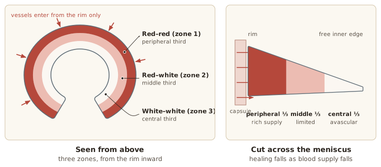
Three zones, from the periphery inward (the slide figure labels them Red Zone aka Zone 1, Red–white zoneThe middle third of the meniscus: limited blood supply and lower healing potential, though repair may still succeed in selected cases. aka Zone 2 and White Zone aka Zone 3):
| Zone | Location | Blood supply and healing | Management |
|---|---|---|---|
| Red–red zoneThe peripheral third of the meniscus: rich capsular blood supply, the greatest potential for biological healing, often suitable for repair. (Zone 1) | Peripheral third | Rich capsular blood supply; greatest potential for biological healing | Often suitable for repair |
| Red–White (Zone 2) | Middle third | Limited blood supply; lower healing potential | Repair may still succeed in selected cases |
| White–white zoneThe central third of the meniscus: avascular, with minimal intrinsic healing, so tears are less likely to heal. (Zone 3) | Central third | AvascularHaving no blood vessels. The inner meniscus is avascular and takes nutrients from the synovial fluid., minimal Intrinsic healingA tissue's own ability to repair itself.General meaning, not from the slides; less likely to heal | SymptomaticCausing symptoms such as pain or swelling.General meaning, not from the slides tears are more often managed by Partial meniscectomySurgery that removes only the unstable or irreparable portion of the meniscus, preserving healthy tissue. |
Zone, tear pattern and patient factors together decide whether a tear is repaired or trimmed.
Nutrition of the avascular meniscus
- No direct blood supply: the inner (avascular) portion has no vessels reaching it.
- DiffusionGradual movement of nutrients from where they are plentiful, here the synovial fluid, into tissue that has no blood vessels. from Synovial fluidThe slippery fluid inside a joint that lubricates it and helps feed the cartilage.General meaning, not from the slides: nutrients are taken up primarily from the synovial fluid.
- Cyclic loading and unloadingRepeated pressing and releasing of the meniscus as the knee moves, which drives exchange of nutrients and waste.: knee movement drives exchange of nutrients and Metabolic wasteWaste products made by cells as they work.General meaning, not from the slides.
- Tissue stays viable: meniscal tissue maintains its function despite avascularity.
Clinical link: tears in the avascular white–white zone depend on diffusion alone and have minimal capacity to heal.
Biomechanics: Seven Functions
The MeniscusOne of two semicircular fibrocartilaginous structures between the femoral condyles and the tibial plateau of the knee: the medial and the lateral meniscus. protect the Articular cartilageThe smooth cartilage that covers the ends of the bones inside a joint.General meaning, not from the slides and keep the knee working normally in seven ways.
| Function | What the menisci do |
|---|---|
| 1 and 2 | Load distribution and Hoop stressCompression pushes the meniscus outward and its circumferential fibers resist by developing tensile force, turning vertical load into circumferential tension. |
| 3 | Shock absorptionSoaking up and spreading out impact forces from walking, running and jumping. |
| 4 | Load sharingThe share of the compressive load carried by the menisci, which rises as the knee bends. during knee motion |
| 5 | Joint stability |
| 6 | LubricationReducing friction between surfaces with a slippery fluid. and nutrition |
| 7 | ProprioceptionThe body's sense of joint position and movement.General meaning, not from the slides |
Load distribution and hoop stress (functions 1 and 2)
- The menisci enlarge the Contact stressThe force spread over the touching surfaces of femur and tibia. A larger contact area means less stress on the cartilage.General meaning, not from the slides between the Femoral condylesThe two rounded knobs at the lower end of the femur that sit on top of the tibia and menisci.General meaning, not from the slides and Tibial plateauThe flat top surface of the tibia on which the femur and the menisci rest.General meaning, not from the slides, spreading load and lowering contact stress on the cartilage.
- They convert vertical CompressionSqueezing together, such as body weight pressing the femur down on the meniscus. In swelling control it also means pressing on the area, for example with a bandage.General meaning, not from the slides into Circumferential fibersCollagen fibers running around the curve of the meniscus. They give tensile strength and resist the outward push of load. Tensile forceA pulling force that stretches a material. Tensile strength is how much pulling it can resist.General meaning, not from the slides (hoop stress), directing force outward to the meniscal attachments.
Without them: faster cartilage DegenerativeCaused by gradual wear and breakdown of tissue over time.General meaning, not from the slides and a higher risk of knee Osteoarthritis (OA)Wear-related joint disease in which cartilage breaks down, causing pain and stiffness.General meaning, not from the slides.
| After Total meniscectomySurgical removal of the entire meniscus.General meaning, not from the slides | Change |
|---|---|
| Tibiofemoral jointThe main knee joint, where the femur meets the tibia.General meaning, not from the slides contact area | ≈50% less |
| Peak contact stress | 200–300% increase |
Shock absorption (function 3)
≈20% drop in the knee’s shock-absorbing capacity after MeniscectomySurgical removal of all or part of a meniscus.General meaning, not from the slides.
- The FibrocartilageTough, springy cartilage reinforced with collagen fibers, able to cope with both squeezing and pulling forces.General meaning, not from the slides dissipates forces generated by walking, running and jumping.
- It absorbs and redistributes impact loads across the joint.
- This shields the articular cartilage and Subchondral boneThe layer of bone just beneath the joint cartilage.General meaning, not from the slides from excess stress.
- After meniscectomy, more impact reaches the joint surfaces.
Load sharing during knee motion (function 4)

FlexionBending the knee.General meaning, not from the slides increases meniscal loading. The share of compressive load carried by the menisci is:
| Knee position | Share of compressive load carried by the menisci |
|---|---|
| Full ExtensionStraightening the knee.General meaning, not from the slides | About 50% |
| 90° flexion | About 85% |
This is why the menisci matter most in squatting, kneeling and stair climbing.
Stability, lubrication and proprioception (functions 5, 6 and 7)
- Function 5, joint stability: better FemurThe thigh bone.General meaning, not from the slides–TibiaThe shin bone, the larger bone of the lower leg.General meaning, not from the slides CongruencyHow well two joint surfaces fit together. A better fit gives a more stable joint. increases stability, especially in flexion and in Ligament-deficient kneeA knee with a torn or missing ligament, for example after ACL injury. such as after ACLAnterior cruciate ligament: a knee ligament that stops the tibia sliding forward on the femur.General meaning, not from the slides injury.
- Function 6, lubrication and nutrition: movement spreads Synovial fluidThe slippery fluid inside a joint that lubricates it and helps feed the cartilage.General meaning, not from the slides over the joint surfaces, reducing friction and helping nutrients reach AvascularHaving no blood vessels. The inner meniscus is avascular and takes nutrients from the synovial fluid. areas of the menisci and cartilage.
- Function 7, proprioception: MechanoreceptorsSensors in the peripheral menisci that sense joint position, movement and load. in the PeripheryThe outer rim of the meniscus, where it attaches to the joint capsule.General meaning, not from the slides menisci sense joint position, movement and load, supporting Neuromuscular controlHow well the nervous system coordinates the muscles to control movement and protect the joint.General meaning, not from the slides and Dynamic stabilityA joint's stability while it is moving, provided by muscle control.General meaning, not from the slides.
Mechanism of Injury
Epidemiology: tears are common and often silent
Framingham cohortThe group of 991 adults older than 50 in the slide's study who had knee MRI regardless of knee pain.: 991 adults older than 50 years who underwent knee MRIMagnetic resonance imaging: a scan that shows soft tissues such as menisci and ligaments.General meaning, not from the slides regardless of knee pain.
| Finding | Result |
|---|---|
| Had MeniscusOne of two semicircular fibrocartilaginous structures between the femoral condyles and the tibial plateau of the knee: the medial and the lateral meniscus. damage on MRI | 35% |
| Of those with Osteoarthritis (OA)Wear-related joint disease in which cartilage breaks down, causing pain and stiffness.General meaning, not from the slides, had meniscal damage | 82% |
| Of tears, were in knees without pain, aching or stiffness | ≈55% |
Clinical message: a meniscal finding on MRI is not automatically the source of symptoms. Always interpret ImagingScans such as MRI that show the inside of the body.General meaning, not from the slides together with the history and examination.
Source on the slide: Englund M, et al. N Engl J Med 2008 (summary: Am Fam Physician 2011, AAFP Journal Club).
Traumatic and degenerative mechanisms
Injuries result from AcuteSudden and recent, as opposed to long-standing.General meaning, not from the slides trauma or DegenerativeCaused by gradual wear and breakdown of tissue over time.General meaning, not from the slides change, depending on age, activity level and the condition of the meniscal tissue.
| Description | |
|---|---|
| Younger individuals | Tears are usually TraumaticCaused by a sudden injury.General meaning, not from the slides and sports related. |
| Older adults | Tears are more commonly degenerative and may occur with minimal trauma. |
| Rises with age | Degeneration reduces the strength and ElasticityThe ability of a tissue to stretch and spring back.General meaning, not from the slides of the FibrocartilageTough, springy cartilage reinforced with collagen fibers, able to cope with both squeezing and pulling forces.General meaning, not from the slides. |
| Medial > lateral | Medial tears are more frequent than lateral tears. |
| Often combined | Frequently associated with ACLAnterior cruciate ligament: a knee ligament that stops the tibia sliding forward on the femur.General meaning, not from the slides, Collateral ligamentA ligament on the inner (medial) or outer (lateral) side of the knee that resists sideways stress.General meaning, not from the slides or Articular cartilageThe smooth cartilage that covers the ends of the bones inside a joint.General meaning, not from the slides injury. |
Acute traumatic tears in younger individuals
Typically occur in sports or other high-demand activities with rapid changes of direction, pivoting or landing.
- Twisting or pivoting: on a Weight bearingPutting body weight through the leg.General meaning, not from the slides knee with the foot firmly planted; commonly produces Longitudinal tearA tear running parallel to the collagen fibers, splitting the meniscus into inner and outer parts. or Bucket-handle tearA longitudinal tear whose displaced fragment causes locking, loss of terminal extension and catching..
- RotationTwisting around an axis.General meaning, not from the slides with FlexionBending the knee.General meaning, not from the slides: sudden rotational forces with knee flexion during cutting, pivoting or changing direction.
- Jump landing: landing with excessive Femorotibial rotationTwisting movement between the femur and the tibia.General meaning, not from the slides or Axial loadingForce pushing straight down along the long axis of the limb.General meaning, not from the slides.
- Contact injury: ValgusAngled so that the lower leg points outward from the thigh, giving a knock-kneed alignment.General meaning, not from the slides or VarusAngled so that the lower leg points inward toward the midline, giving a bow-legged alignment.General meaning, not from the slides stress with rotation or HyperextensionBending a joint backward beyond its normal straight position.General meaning, not from the slides; may cause associated ligament injuries.
Acute injury vs degenerative change
| Acute injury / trauma | Degenerative change |
|---|---|
| Often occurs during activities involving twisting or pivoting of the knee | Wear and tear over time weakens the meniscus, leading to tears |
Clinical Assessment
Clinical presentation of a meniscal tear
Pain, swelling and Mechanical symptomsLocking, clicking and grinding of the knee. are typical. Severity depends on the type, size and location of the tear.
- Joint line tendernessLocalised tenderness along the medial or lateral joint line, one of the most reliable findings of a meniscal tear.: localised TendernessPain or discomfort when a body part is pressed.General meaning, not from the slides along the medial or lateral joint line is one of the most reliable findings. Best assessed with the knee flexed to 45–90°.
- Joint effusionExtra fluid in the joint that causes swelling.General meaning, not from the slides: mild to moderate swelling may develop within 24–48 hours of injury. It is common, but its absence does not exclude a tear.
- Pain: localised to the affected joint line; aggravated by deep squatting, kneeling, twisting, pivoting or stairs. Horn of the meniscusThe front (anterior horn) or back (posterior horn) end of the crescent-shaped meniscus.General meaning, not from the slides tears hurt in deep FlexionBending the knee.General meaning, not from the slides.
- Mechanical symptoms: LockingTrue knee locking: a displaced meniscal fragment becomes trapped and mechanically prevents full extension., ClickingA snapping sound or sensation in the joint during movement.General meaning, not from the slides, GrindingA rough, gritty feeling or sound in the joint during movement.General meaning, not from the slides.
True knee locking
- What it is: a DisplacedMoved out of its normal position.General meaning, not from the slides MeniscusOne of two semicircular fibrocartilaginous structures between the femoral condyles and the tibial plateau of the knee: the medial and the lateral meniscus. fragment, most commonly from a Bucket-handle tearA longitudinal tear whose displaced fragment causes locking, loss of terminal extension and catching., becomes trapped between FemurThe thigh bone.General meaning, not from the slides and TibiaThe shin bone, the larger bone of the lower leg.General meaning, not from the slides and mechanically prevents full ExtensionStraightening the knee.General meaning, not from the slides.
- How it feels on examination: a firm or springy mechanical block to extension, rather than pain-limited motion.
- Prompt OrthopedicRelating to bones, joints and muscles.General meaning, not from the slides evaluation: a displaced tear often needs surgery.

The slide photo shows a model knee with a displaced meniscus (caught in the joint) that blocks the knee from straightening; the inset compares a normal knee (meniscus in normal position) with a locked knee (torn meniscus flipped into the joint causing a block).
Physical examination
No single test confirms or excludes a meniscal tear. Combine history, clinical findings and ImagingScans such as MRI that show the inside of the body.General meaning, not from the slides when indicated. The more findings are present, the higher the likelihood of a tear.
| Step | Findings |
|---|---|
| History | Twisting or pivoting injury to a Weight bearingPutting body weight through the leg.General meaning, not from the slides knee; locking, CatchingA sensation of the knee snagging during movement.General meaning, not from the slides or clicking |
| InspectionExamining by looking, for example for swelling or muscle wasting.General meaning, not from the slides | Quadriceps atrophyShrinking and weakening of the quadriceps muscle. The slide says it is more common in chronic injury. (check the VMOVastus medialis obliquus: the lowest, inner part of the quadriceps, on the inner side of the knee.General meaning, not from the slides; more common in ChronicLong-standing, as opposed to recent.General meaning, not from the slides injury). Effusion (may appear 24–48 h after injury; often absent in small tears). Sweep (brush) testA test that assesses effusion in the knee. assesses effusion |
| PalpationExamining by feeling with the hands.General meaning, not from the slides and motion | Localised joint line tenderness; pain on forced HyperextensionBending a joint backward beyond its normal straight position.General meaning, not from the slides (Modified bounce home testPain on forced hyperextension of the knee, which raises suspicion of a meniscal tear.); pain on Maximal passive flexionThe examiner bends the knee as far as it goes while the patient relaxes. Pain on this movement raises suspicion of a tear. |
| Provocative testsTests that try to reproduce the patient's symptoms.General meaning, not from the slides | McMurray testReproduces pain, clicking or a palpable clunk during tibiofemoral rotation while the knee is flexed and extended.: pain or palpable/audible click. Thessaly testA dynamic weight-bearing test that reproduces joint line pain or mechanical symptoms during rotational loading.: positive |
Findings that raise suspicion of a tear
Slide 28 repeats the examination findings as a checklist:
- Twisting or pivoting injury to a weight-bearing knee
- Locking, catching or clicking
- Localised joint line tenderness
- Pain on forced hyperextension (modified bounce home test)
- Pain on maximal passive flexion
- McMurray test: pain or palpable/audible click
- Positive Thessaly test
Combine findings: no single sign is diagnostic. Next step: confirm with special tests, and with imaging when it would change management.
Palpation: joint line tenderness
- 45–90° of knee flexion exposes the joint line for palpation of the medial and lateral sides.
- Why it matters: palpation of the medial and lateral joint lines is one of the most valuable components of the Physical examinationThe hands-on examination of the patient by the clinician.General meaning, not from the slides.
- Interpretation: localised tenderness over the involved meniscus is highly suggestive of a tear, particularly with an appropriate injury mechanism.
Slides 25 and 29 call joint line tenderness one of the most reliable or most valuable findings and highly suggestive of a tear, while slides 27, 28 and 31 say no single test or sign is diagnostic. Both are on the slides: use joint line tenderness together with the other findings.
Special tests for meniscal injury
These tests apply CompressionSqueezing together, such as body weight pressing the femur down on the meniscus. In swelling control it also means pressing on the area, for example with a bandage.General meaning, not from the slides, RotationTwisting around an axis.General meaning, not from the slides or stress to the meniscus to reproduce pain, clicking, locking or joint line tenderness.
| Test | Purpose and interpretation |
|---|---|
| McMurray test | Reproduces pain, clicking or a palpable ClunkA dull knock that can be felt or heard in the joint.General meaning, not from the slides during Femorotibial rotationTwisting movement between the femur and the tibia.General meaning, not from the slides while flexing and extending the knee. |
| Thessaly test | Dynamic weight-bearing test that reproduces joint line pain or mechanical symptoms during rotational loading. |
| Joint line tenderness | Assesses localised tenderness along the medial or lateral joint line; most useful when combined with other findings. |
| Apley compression testAxial compression with tibial rotation, patient prone. Pain on compression suggests a meniscal lesion, pain on distraction suggests ligament injury. | Axial loadingForce pushing straight down along the long axis of the limb.General meaning, not from the slides with tibial rotation, patient ProneLying face down.General meaning, not from the slides. Pain on compression suggests a meniscal LesionAn area of damaged tissue.General meaning, not from the slides; pain on DistractionGently pulling the joint surfaces apart.General meaning, not from the slides suggests ligament injury. |
How accurate are the clinical tests?

Meta-analysisA study that combines the results of several earlier studies.General meaning, not from the slides diagnostic accuracy (%), from a Systematic reviewA structured review that gathers and summarises all the studies on a question.General meaning, not from the slides and meta-analysis:
| Test | SensitivityHow well a test picks up people who truly have the condition.General meaning, not from the slides | SpecificityHow well a test correctly clears people who do not have the condition.General meaning, not from the slides |
|---|---|---|
| Joint line tenderness | 83% | 83% |
| McMurray | 61% | 84% |
| Thessaly (20°) | 75% | 87% |
- Joint line tenderness was the most sensitive (83%). Thessaly at 20° was the most specific (87%).
- The review authors found the evidence HeterogeneousMade up of differing parts, here studies that differ from one another.General meaning, not from the slides and low quality, and concluded that none of these tests alone can be recommended for diagnosis. Use clusters of findings.
Source on the slide: Smith BE, et al. Evid Based Med 2015.
Stoller MRI grading of meniscal degeneration
| Grade | MRIMagnetic resonance imaging: a scan that shows soft tissues such as menisci and ligaments.General meaning, not from the slides findings | Clinical significance |
|---|---|---|
| Grade 1 | Small focal Intrameniscal signalA bright area seen inside the meniscus on MRI.General meaning, not from the slides that does not reach an Articular surfaceThe smooth, cartilage-covered surface of a bone inside the joint.General meaning, not from the slides | IntrasubstanceWithin the substance of the meniscus, without reaching its surface.General meaning, not from the slides DegenerativeCaused by gradual wear and breakdown of tissue over time.General meaning, not from the slides; not a true tear |
| Grade 2 | Linear intrameniscal signal that does not extend to the articular surface | Degenerative change; generally not a tear |
| Grade 3 | Signal extends to one or both articular surfaces | True meniscal tear |
Grades 1 and 2 are common with aging and often AsymptomaticCausing no symptoms.General meaning, not from the slides: interpret them with the history and examination. Only Grade 3 is a true tear on MRI.
Supporting evidence: in adults older than 50, MRI meniscal damage was found in 35% and about 55% of tears were asymptomatic (Englund 2008).
Classification of Tears
Tear pattern, location and VascularHaving a blood supply.General meaning, not from the slides zone guide whether a tear can be repaired.

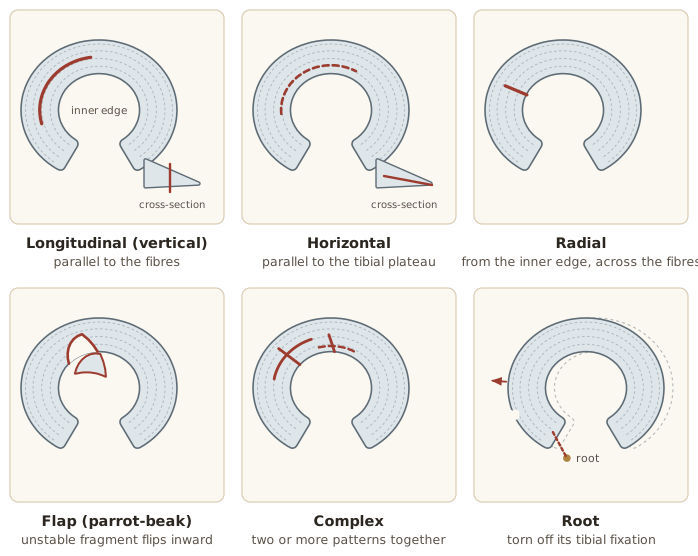
The slide shows two sets of figures: a labelled illustration of the knee with six tear types (IntrasubstanceWithin the substance of the meniscus, without reaching its surface.General meaning, not from the slides, Horizontal tearA tear running parallel to the tibial plateau, splitting the meniscus into superior and inferior parts., Radial tearA tear starting at the free inner edge and extending toward the peripheral rim, crossing the collagen fibers., Flap tearA tear arising from a horizontal or oblique tear that leaves an unstable fragment., Bucket-handle tearA longitudinal tear whose displaced fragment causes locking, loss of terminal extension and catching., Complex tearA tear combining two or more tear patterns (longitudinal, horizontal, radial or flap).) and a panel of MeniscusOne of two semicircular fibrocartilaginous structures between the femoral condyles and the tibial plateau of the knee: the medial and the lateral meniscus. outlines labelled normal, vertical Longitudinal tearA tear running parallel to the collagen fibers, splitting the meniscus into inner and outer parts., vertical radial, horizontal, Oblique tearA tear running at a slant across the meniscus.General meaning, not from the slides, complex/DegenerativeCaused by gradual wear and breakdown of tissue over time.General meaning, not from the slides.
A. Longitudinal (vertical)
Runs parallel to the CollagenThe main structural protein of tendons, ligaments and cartilage.General meaning, not from the slides fibers, splitting the meniscus into inner and outer parts.
- Usually TraumaticCaused by a sudden injury.General meaning, not from the slides; common in young athletes.
- Often in the vascular PeripheryThe outer rim of the meniscus, where it attaches to the joint capsule.General meaning, not from the slides; heals well if repaired early.
- Bucket-handle tear: DisplacedMoved out of its normal position.General meaning, not from the slides fragment causes LockingTrue knee locking: a displaced meniscal fragment becomes trapped and mechanically prevents full extension., loss of Terminal extensionThe last few degrees of straightening the knee.General meaning, not from the slides, CatchingA sensation of the knee snagging during movement.General meaning, not from the slides.
B. Horizontal
Runs parallel to the Tibial plateauThe flat top surface of the tibia on which the femur and the menisci rest.General meaning, not from the slides, splitting the meniscus into superior and inferior parts.
- Common in degenerative menisci.
- Seen in middle-aged and older adults.
- May communicate with Meniscal cystA fluid-filled swelling next to a meniscus.General meaning, not from the slides and extend obliquely.
C. Radial
Starts at the Free edgeThe thin inner border of the meniscus that faces the middle of the joint.General meaning, not from the slides and extends toward the peripheral rim, crossing the collagen fibers.
- Disrupts Hoop stressCompression pushes the meniscus outward and its circumferential fibers resist by developing tensile force, turning vertical load into circumferential tension. and raises Tibiofemoral jointThe main knee joint, where the femur meets the tibia.General meaning, not from the slides Contact stressThe force spread over the touching surfaces of femur and tibia. A larger contact area means less stress on the cartilage.General meaning, not from the slides.
- May cause meniscal Meniscal extrusionThe meniscus being pushed out from its normal position at the edge of the joint.General meaning, not from the slides.
- Large tears act like MeniscectomySurgical removal of all or part of a meniscus.General meaning, not from the slides; Meniscal repairSurgery that preserves meniscal function and lowers long-term osteoarthritis risk. It is the preferred surgical option on the slides. whenever feasible.
D. Flap (parrot-beak)
Arises from a horizontal or oblique tear, leaving an unstable fragment.
- Causes intermittent pain and Mechanical symptomsLocking, clicking and grinding of the knee..
- Fragment may catch between FemurThe thigh bone.General meaning, not from the slides and TibiaThe shin bone, the larger bone of the lower leg.General meaning, not from the slides.
- Mostly degenerative, but can follow trauma.
E. Complex
Combines two or more tear patterns (longitudinal, horizontal, radial or flap).
- Most common in degenerative menisci.
- Frequently associated with Osteoarthritis (OA)Wear-related joint disease in which cartilage breaks down, causing pain and stiffness.General meaning, not from the slides.
- Extensive tissue involvement; repair is less successful.
F. Root
Disrupts the anterior or posterior Meniscal rootThe anterior or posterior end where the meniscus is firmly anchored to the tibial plateau. Root attachments fix each horn to the tibia., so the meniscus loses its fixation to the tibia.
- Most often the posterior root of the medial meniscus.
- Causes extrusion; effects comparable to Total meniscectomySurgical removal of the entire meniscus.General meaning, not from the slides.
- Early repair reduces rapid Articular cartilageThe smooth cartilage that covers the ends of the bones inside a joint.General meaning, not from the slides degeneration and OA.
Comparison of the six patterns
| Pattern | Direction | Typical setting | Other key points on the slide |
|---|---|---|---|
| A. Longitudinal | Parallel to collagen fibers (inner vs outer parts) | Traumatic, young athletes | Heals well if repaired early; bucket-handle form causes locking |
| B. Horizontal | Parallel to tibial plateau (superior vs inferior parts) | Degenerative, middle-aged and older adults | May communicate with meniscal cysts and extend obliquely |
| C. Radial | From free inner edge toward peripheral rim | Not stated | Disrupts hoop stress; repair whenever feasible (large tears act like meniscectomy) |
| D. Flap | From a horizontal or oblique tear | Mostly degenerative, can follow trauma | Unstable fragment, intermittent pain and mechanical symptoms |
| E. Complex | Two or more patterns combined | Most common in degenerative menisci | Frequently associated with OA; repair is less successful |
| F. Root | Anterior or posterior root | Most often posterior root of the medial meniscus | Causes extrusion; early repair reduces rapid cartilage degeneration and OA |
Slide 35 says horizontal tears are “common” in degenerative menisci, while slide 36 says complex tears are “most common” in degenerative menisci. Both wordings are kept as written.
Management
Meniscal preservation: healing by zone
| Zone | Healing potential | What it means |
|---|---|---|
| Red–red zoneThe peripheral third of the meniscus: rich capsular blood supply, the greatest potential for biological healing, often suitable for repair. | Highest healing potential | Usually suitable for Meniscal repairSurgery that preserves meniscal function and lowers long-term osteoarthritis risk. It is the preferred surgical option on the slides. |
| Red–white zoneThe middle third of the meniscus: limited blood supply and lower healing potential, though repair may still succeed in selected cases. | May heal successfully | Depends on tear characteristics and patient factors |
| White–white zoneThe central third of the meniscus: avascular, with minimal intrinsic healing, so tears are less likely to heal. | Limited healing potential | Poor VascularHaving a blood supply.General meaning, not from the slides; more likely to need Partial meniscectomySurgery that removes only the unstable or irreparable portion of the meniscus, preserving healthy tissue. if SymptomaticCausing symptoms such as pain or swelling.General meaning, not from the slides |
Why preserve: healthy MeniscusOne of two semicircular fibrocartilaginous structures between the femoral condyles and the tibial plateau of the knee: the medial and the lateral meniscus. tissue maintains normal knee BiomechanicsHow forces act on and through the body during movement.General meaning, not from the slides, reduces Contact stressThe force spread over the touching surfaces of femur and tibia. A larger contact area means less stress on the cartilage.General meaning, not from the slides and lowers the long-term risk of Articular cartilageThe smooth cartilage that covers the ends of the bones inside a joint.General meaning, not from the slides DegenerativeCaused by gradual wear and breakdown of tissue over time.General meaning, not from the slides and Osteoarthritis (OA)Wear-related joint disease in which cartilage breaks down, causing pain and stiffness.General meaning, not from the slides.
Clinical pearl: repair over meniscectomy
Repair over MeniscectomySurgical removal of all or part of a meniscus.General meaning, not from the slides for repairable tears.
- Best candidates for repair: Longitudinal tearA tear running parallel to the collagen fibers, splitting the meniscus into inner and outer parts., Bucket-handle tearA longitudinal tear whose displaced fragment causes locking, loss of terminal extension and catching., Radial tearA tear starting at the free inner edge and extending toward the peripheral rim, crossing the collagen fibers. and Root tearA tear that disrupts the anterior or posterior root, so the meniscus loses its fixation to the tibia. in vascular regions.
- Why preserve tissue: better long-term knee function and a lower risk of osteoarthritis.
Conservative management
Best suited for:
- Small, stable tears
- Tears in the vascular red–red or red–white zones
- Degenerative tears without significant Mechanical symptomsLocking, clicking and grinding of the knee.
- Minimal pain and preserved knee function
- Patients who are not surgical candidates
Structured program:
- Education and activity modification
- Short-term pain and swelling control
- Progressive Physical therapyTreatment with exercise and hands-on techniques to restore movement and function.General meaning, not from the slides; restore Range of motionHow far a joint can move.General meaning, not from the slides
- QuadricepsThe large muscle group at the front of the thigh.General meaning, not from the slides, HamstringThe muscle group at the back of the thigh.General meaning, not from the slides and hip strengthening
- Neuromuscular controlHow well the nervous system coordinates the muscles to control movement and protect the joint.General meaning, not from the slides and ProprioceptionThe body's sense of joint position and movement.General meaning, not from the slides training
- Gradual return to functional and sporting activities
Many degenerative tears improve with structured RehabilitationExercise and treatment that restore function after injury or surgery.General meaning, not from the slides and do not require surgery.
Surgical management
Considered when Conservative managementTreatment without surgery.General meaning, not from the slides fails or the tear is unlikely to heal without surgery.
| Option | Description | Indications |
|---|---|---|
| Meniscal repair (preferred) | Preserves meniscal function and lowers long-term osteoarthritis risk | Longitudinal, bucket-handle, repairable radial and root tears; red–red or red–white zones; AcuteSudden and recent, as opposed to long-standing.General meaning, not from the slides TraumaticCaused by a sudden injury.General meaning, not from the slides tears; young or active patients; tears with ACLAnterior cruciate ligament: a knee ligament that stops the tibia sliding forward on the femur.General meaning, not from the slides ReconstructionSurgery that rebuilds a torn ligament, for example the ACL.General meaning, not from the slides |
| Partial meniscectomy | Removes only the unstable or irreparable portion, preserving healthy tissue | Irreparable or severely degenerative tears; Complex tearA tear combining two or more tear patterns (longitudinal, horizontal, radial or flap).; white–white zone; persistent mechanical symptoms from unstable fragments |
| DebridementSmoothing of frayed or unstable tissue while keeping the functional meniscus. | Smooths frayed or unstable tissue while keeping the functional meniscus | Commonly performed during arthroscopic partial meniscectomy |
| Allograft transplantationTransplanting donor meniscus tissue, which restores load distribution, reduces pain and delays osteoarthritis. | Restores load distribution, reduces pain and delays osteoarthritis | Carefully selected young patients after Subtotal meniscectomySurgical removal of most, but not all, of the meniscus.General meaning, not from the slides or Total meniscectomySurgical removal of the entire meniscus.General meaning, not from the slides with persistent CompartmentA section of the knee joint, such as the medial (inner) or lateral (outer) side.General meaning, not from the slides pain and minimal osteoarthritis |
Routine arthroscopic partial meniscectomy is discouraged for degenerative tears without mechanical LockingTrue knee locking: a displaced meniscal fragment becomes trapped and mechanically prevents full extension..
Indications for surgical intervention
- Persistent mechanical locking from a DisplacedMoved out of its normal position.General meaning, not from the slides meniscal fragment
- Symptomatic bucket-handle or root tears
- Large, unstable traumatic tears
- Tears with ACL RuptureA complete tear or break of a tissue such as a ligament.General meaning, not from the slides or other ligament injury needing reconstruction
- Persistent pain, swelling or limitation despite 3–6 months of conservative care
- Young or highly active patients who need normal knee biomechanics restored
Evidence: degenerative tears, surgery vs sham (FIDELITY trial)
- Randomised, double-blindPatients are assigned to treatments by chance (randomised), and neither patients nor assessors know who got which treatment (double-blind).General meaning, not from the slides, double-blind, Sham surgeryA pretend operation, used in trials to test whether the real operation works.General meaning, not from the slides-controlled (Finland).
- 146 patients aged 35–65 with a degenerative medial meniscus tear and no osteoarthritis.
- Arthroscopic partial meniscectomy vs sham surgery.
- Follow-up: 12 months.

Improvement from baseline at 12 months (points):
| Score | Partial meniscectomy | Sham surgery |
|---|---|---|
| Lysholm scoreA questionnaire score of knee function and symptoms.General meaning, not from the slides | 22 | 23 |
| WOMETWestern Ontario Meniscal Evaluation Tool: a questionnaire score for symptoms from a meniscal problem.General meaning, not from the slides | 25 | 27 |
No significant between-group differences in Lysholm, WOMET or knee pain after exercise. Outcomes after partial meniscectomy were no better than after sham surgery.
Source on the slide: Sihvonen R, et al. N Engl J Med 2013.
Clinical pearl: match treatment to tear type
| Tear type | Treatment |
|---|---|
| Degenerative tears | Exercise-based rehabilitation is first-line treatment. Reserve ArthroscopicDone through small cuts using a camera and instruments placed inside the joint.General meaning, not from the slides surgery for persistent disabling symptoms or true mechanical locking. |
| Acute traumatic tears in young, active patients | Repair whenever feasible, especially longitudinal, bucket-handle, radial and root tears, to preserve meniscal function and long-term joint health. |
Rehabilitation and Return to Sport
Rehabilitation considerations
RehabilitationExercise and treatment that restore function after injury or surgery.General meaning, not from the slides depends on:
- Type of injury
- Treatment approach (Conservative managementTreatment without surgery.General meaning, not from the slides or surgical)
- Healing status of the MeniscusOne of two semicircular fibrocartilaginous structures between the femoral condyles and the tibial plateau of the knee: the medial and the lateral meniscus.
Primary goals:
- Reduce pain and swelling
- Restore knee function
- Regain muscle strength
- Return safely to daily activities and sport while protecting the healing meniscus
Three pathways: Non-operativeWithout surgery.General meaning, not from the slides, after Partial meniscectomySurgery that removes only the unstable or irreparable portion of the meniscus, preserving healthy tissue., and after Meniscal repairSurgery that preserves meniscal function and lowers long-term osteoarthritis risk. It is the preferred surgical option on the slides..
Pathway 1: non-operative rehabilitation
For stable or DegenerativeCaused by gradual wear and breakdown of tissue over time.General meaning, not from the slides tears without significant Mechanical symptomsLocking, clicking and grinding of the knee..
Rehabilitation goals:
- Control pain and joint swelling
- Restore full knee Range of motionHow far a joint can move.General meaning, not from the slides
- Maintain and build QuadricepsThe large muscle group at the front of the thigh.General meaning, not from the slides and lower-limb strength
- Improve Neuromuscular controlHow well the nervous system coordinates the muscles to control movement and protect the joint.General meaning, not from the slides, balance and ProprioceptionThe body's sense of joint position and movement.General meaning, not from the slides
- Return gradually to functional and sporting activities
Avoid early on:
- Deep squatting
- Pivoting and twisting
- Cutting movements
- Running and jumping
Resume the avoided activities gradually as symptoms and function improve.
Pathway 2: after partial meniscectomy
| Principle | Guidance |
|---|---|
| Weight bearingPutting body weight through the leg.General meaning, not from the slides | Usually allowed immediately as tolerated |
| Range of motion | Early full range of motion is encouraged |
| Strengthening | Begin quadriceps activation and progressive strengthening as soon as tolerated |
| Swelling control | CryotherapyTreatment with cold, such as ice.General meaning, not from the slides, CompressionSqueezing together, such as body weight pressing the femur down on the meniscus. In swelling control it also means pressing on the area, for example with a bandage.General meaning, not from the slides, ElevationRaising the limb to help reduce swelling.General meaning, not from the slides and activity modification |
| Return to activity | Most patients resume normal daily activities within 2–6 weeks |
Recovery is generally rapid because no meniscal healing is required. Routine ImmobilisationKeeping a joint still, for example in a brace.General meaning, not from the slides is not recommended unless the surgeon specifies it.
Pathway 3: after meniscal repair
Weight bearing
| Stage | Timing | Guidance |
|---|---|---|
| 1 | Immediately after surgery | Brace commonly used, locked in full ExtensionStraightening the knee.General meaning, not from the slides for walking to protect the repair |
| 2 | First 4–6 weeks | Weight bearing limited or progressed gradually according to the repair and the surgeon’s ProtocolThe surgeon's step-by-step plan for recovery after surgery.General meaning, not from the slides |
| 3 | After 6 weeks | Advance to full weight bearing as tolerated once healing and quadriceps control are adequate |
| 4 | Complex tearA tear combining two or more tear patterns (longitudinal, horizontal, radial or flap). or Root tearA tear that disrupts the anterior or posterior root, so the meniscus loses its fixation to the tibia. | Restrictions may extend to 6–8 weeks to protect the healing tissue |
Follow the surgeon’s protocol: restrictions vary with tear pattern, repair technique, associated procedures (e.g., ACLAnterior cruciate ligament: a knee ligament that stops the tibia sliding forward on the femur.General meaning, not from the slides ReconstructionSurgery that rebuilds a torn ligament, for example the ACL.General meaning, not from the slides) and any IntraoperativeHappening during an operation.General meaning, not from the slides complications.
Range of motion
Early controlled motion is encouraged; prolonged immobilisation impairs meniscal nutrition and CollagenThe main structural protein of tendons, ligaments and cartilage.General meaning, not from the slides organisation.
- Restore extension gradually
- Progress FlexionBending the knee.General meaning, not from the slides in a controlled manner
- Avoid loaded flexion beyond 90° for the first 6–8 weeks
- Delay deep squatting and kneeling until healed
Two different time frames appear here: weight bearing (limited for the first 4–6 weeks, up to 6–8 weeks for complex or root repairs) and loaded flexion beyond 90° (avoid for the first 6–8 weeks).
Strengthening progression
- Quadriceps activation and strengthening
- Closed-chain exercisesExercises in which the foot stays fixed on the ground or a surface.General meaning, not from the slides within permitted range
- Balance and proprioceptive training
- Functional movement training
- Sport-specific rehabilitation before Return to sport (RTS)Going back safely to training and competition, judged using return-to-sport criteria.
Limb alignment considerations
| Effect | |
|---|---|
| VarusAngled so that the lower leg points inward toward the midline, giving a bow-legged alignment.General meaning, not from the slides Limb alignmentHow the bones of the leg line up. Varus and valgus are two kinds of abnormal alignment.General meaning, not from the slides | Increases loading on the medial CompartmentA section of the knee joint, such as the medial (inner) or lateral (outer) side.General meaning, not from the slides and medial meniscus |
| ValgusAngled so that the lower leg points outward from the thigh, giving a knock-kneed alignment.General meaning, not from the slides alignment | Increases loading on the lateral compartment and lateral meniscus |
| Clinical impact | Abnormal alignment may increase stress on the repaired meniscus, delay healing and contribute to recurrent symptoms |
| Option | In selected patients, an Unloader braceA brace that may reduce loading on one side of the knee during rehabilitation. may reduce compartmental loading during rehabilitation |
Return to sport after meniscal repair
| Measure | Value |
|---|---|
| Mean time to return to running | ≈12.9 weeks |
| Mean time to return to sport | ≈20 weeks (about 5 months) |
| Reported return-to-sport rates | 80–100% |
| Criteria | Requirement |
|---|---|
| Strength | Quadriceps and HamstringThe muscle group at the back of the thigh.General meaning, not from the slides DeficitA shortfall, here in strength compared with the uninjured side.General meaning, not from the slides vs the uninjured side of no more than 30% for return to running and no more than 15% for return to sport |
| Clinical | Full range of motion, no pain, no Quadriceps atrophyShrinking and weakening of the quadriceps muscle. The slide says it is more common in chronic injury. and no Joint effusionExtra fluid in the joint that causes swelling.General meaning, not from the slides |
| Performance | Satisfactory proprioception, balance and neuromuscular tests |
Caveat: criteria across studies are HeterogeneousMade up of differing parts, here studies that differ from one another.General meaning, not from the slides and the evidence is low level (Level IV evidenceA low level of evidence, as the slide describes the return-to-sport criteria.). Individualise decisions and follow the surgeon’s protocol.
Source on the slide: Schwach M, et al. Orthop Traumatol Surg Res 2023 (Scoping reviewA review that maps the research available on a topic.General meaning, not from the slides of return-to-sport criteria after isolated meniscus SutureA stitch, or the repair of tissue with stitches.General meaning, not from the slides).
Key Takeaway and Sources
Protect the MeniscusOne of two semicircular fibrocartilaginous structures between the femoral condyles and the tibial plateau of the knee: the medial and the lateral meniscus., protect the Articular cartilageThe smooth cartilage that covers the ends of the bones inside a joint.General meaning, not from the slides. Losing meniscal tissue exposes the cartilage to higher stress and raises the risk of Osteoarthritis (OA)Wear-related joint disease in which cartilage breaks down, causing pain and stiffness.General meaning, not from the slides. The menisci spread load, absorb Shock absorptionSoaking up and spreading out impact forces from walking, running and jumping., and stabilise and lubricate.
Sources cited on the closing slide:
- Englund M, et al. Incidental meniscal findings on knee MRIMagnetic resonance imaging: a scan that shows soft tissues such as menisci and ligaments.General meaning, not from the slides in middle-aged and elderly persons. N Engl J Med 2008.
- Summary of Englund 2008. Am Fam Physician 2011 (AAFP Journal Club).
- Sihvonen R, et al. Partial meniscectomySurgery that removes only the unstable or irreparable portion of the meniscus, preserving healthy tissue. versus Sham surgeryA pretend operation, used in trials to test whether the real operation works.General meaning, not from the slides for a DegenerativeCaused by gradual wear and breakdown of tissue over time.General meaning, not from the slides meniscal tear. N Engl J Med 2013.
- Smith BE, et al. Special tests for assessing meniscal tears within the knee: a Systematic reviewA structured review that gathers and summarises all the studies on a question.General meaning, not from the slides and Meta-analysisA study that combines the results of several earlier studies.General meaning, not from the slides. Evid Based Med 2015.
- Schwach M, et al. Return to sport (RTS)Going back safely to training and competition, judged using return-to-sport criteria. criteria after isolated meniscus SutureA stitch, or the repair of tissue with stitches.General meaning, not from the slides: Scoping reviewA review that maps the research available on a topic.General meaning, not from the slides of the literature. Orthop Traumatol Surg Res 2023.
The slide states that the original lecture content, images and Stoller gradingA 3-grade MRI scale for meniscal signal changes. Only Grade 3 is a true tear. are by Prof. Dr Akram Fathy, and that the slides added in this version are: objectives and roadmap, EpidemiologyThe study of how common a condition is and who it affects.General meaning, not from the slides, test accuracy, FIDELITY trialA randomised, double-blind, sham-controlled trial from Finland: arthroscopic partial meniscectomy versus sham surgery in 146 patients. evidence and return-to-sport criteria.