Rehabilitation of Ligamentous Knee Injuries (ACL 2) and PCL Rehab
Quiz this lecture (110 questions)Prof. Dr. Nasr Awad’s lecture (PT 406, Physical Therapy for Sport Injuries) is about the PCLPosterior cruciate ligament: runs from the medial femoral condyle inside the notch to the back of the tibial plateau. It is the primary restraint to the tibia sliding backwards. (PCL): its anatomy and function, the Mechanism of injuryHow an injury happens: the force, direction and position of the limb when it occurs.General meaning, not from the slides, clinical evaluation and biomechanics, grading, imaging, non-surgical care and indications for surgery, and the PCL rehabilitation protocol, ending with the criteria for return to sports.
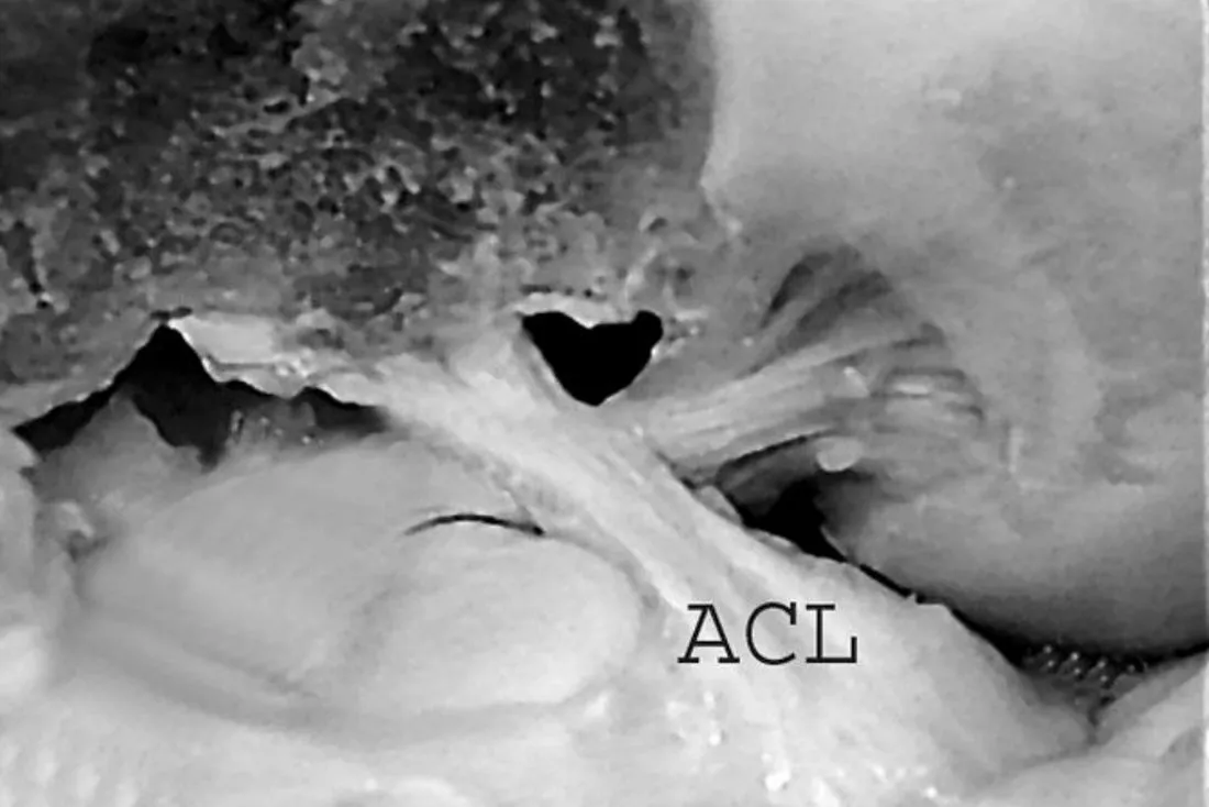
The title slide mentions ACLAnterior cruciate ligament: the knee ligament that mainly stops the tibia sliding forward on the femur.General meaning, not from the slides (“ACL_2”) as well as “PCL rehab”, but every content slide in this file is about the PCL. The ACL appears only in comparisons (the Lachman testAn ACL test: with the knee slightly bent, the examiner pulls the tibia forward to see how far it moves.General meaning, not from the slides warning, healing compared with the ACL) and in one photo labelled ACL.
Objectives
By the end of the lecture, the student should be able to:
- Understand the basic anatomy, function and biomechanics of the PCLPosterior cruciate ligament: runs from the medial femoral condyle inside the notch to the back of the tibial plateau. It is the primary restraint to the tibia sliding backwards..
- Explain the Mechanism of injuryHow an injury happens: the force, direction and position of the limb when it occurs.General meaning, not from the slides of the PCL.
- Discuss PCL risk injuries and how to prevent them.
- Discuss the findings of Stress testA test that applies a force to a joint to check how stable its ligaments are.General meaning, not from the slides for the CruciateMeans 'cross-shaped'. The cruciate ligaments are the two ligaments, anterior and posterior, that cross inside the knee.General meaning, not from the slides injuries.
- Differentiate between OperativeInvolving surgery.General meaning, not from the slides and non-operative treatment of PCL injuries.
- Formulate a proper plan of treatment based on patient presentation and stage of operation.
Objective 3 mentions prevention, but no slide in this file covers it.
PCL Anatomy
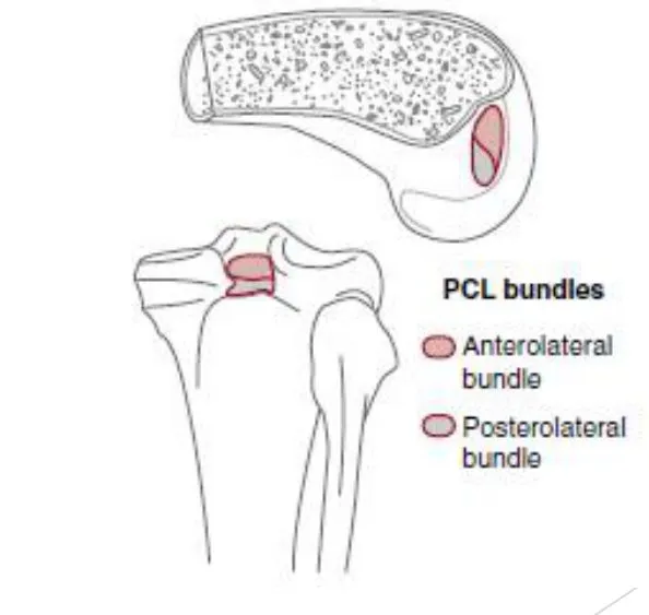
The two bundles
| Larger BundleOne of the separate groups of fibres that together make up the PCL. | Smaller bundle | |
|---|---|---|
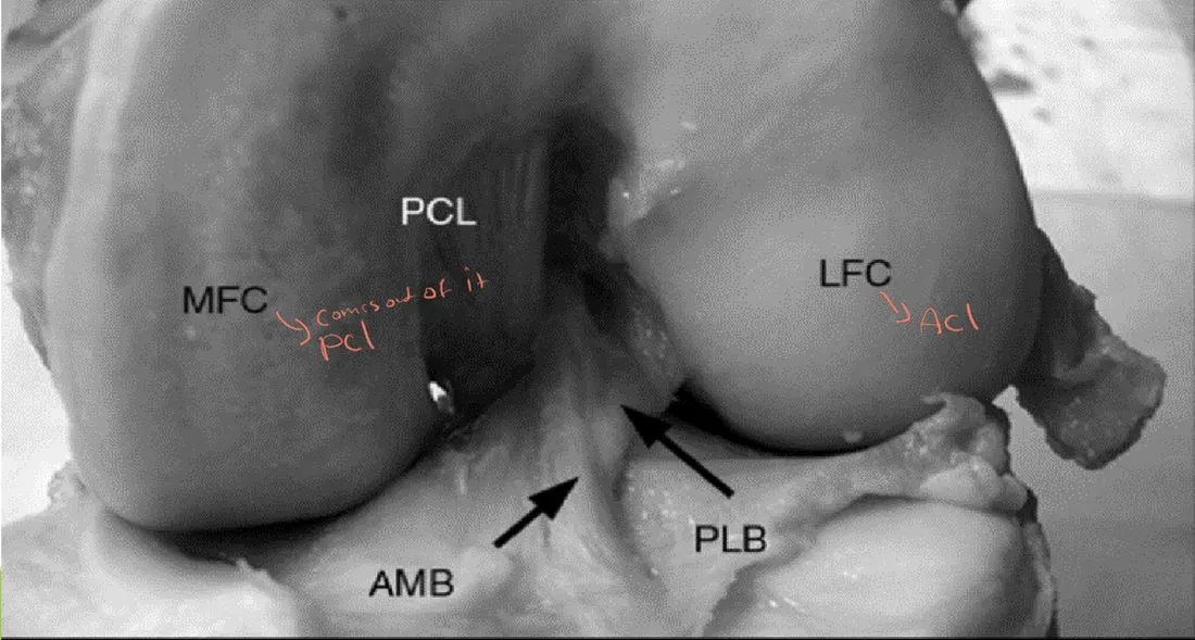
| Name on the bundle slide | Anteromedial bundle (AMB)The larger PCL bundle on the bundle slide, tense in flexion. Later slides call the larger bundle the anterolateral bundle. | Posterolateral bundle (PLB)The smaller PCL bundle on the bundle slide, tense in extension. |
| Size | LARGER | SMALLER |
| Tension | Tension in FlexionBending a joint, for example bending the knee.General meaning, not from the slides | Tension in ExtensionStraightening a joint, for example straightening the knee.General meaning, not from the slides |
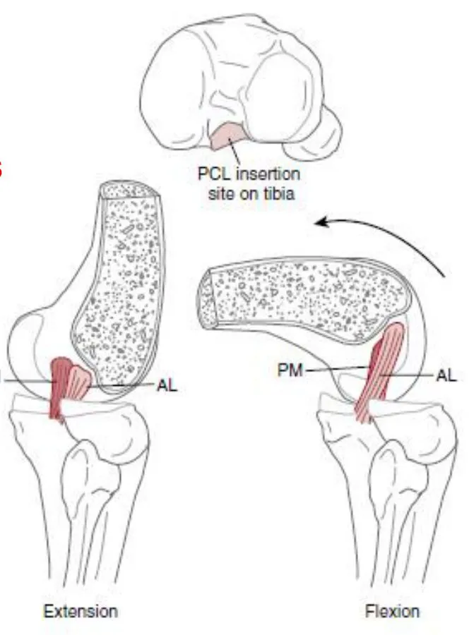
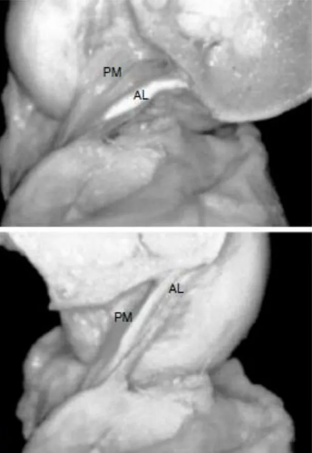
- At its MidsubstanceThe middle part of a ligament, away from its bony attachments.General meaning, not from the slides, the Anterolateral bundle (AL)The larger PCL bundle in the later slides: about twice the size of the posteromedial bundle, stiffer, with a higher ultimate load to failure, and progressively tensed in flexion. is approximately twice the size of the Posteromedial bundle (PM)The smaller PCL bundle in the later slides, compared with the anterolateral bundle. in Cross-sectionThe view you get by cutting straight across a structure, used here to compare how thick the two bundles are.General meaning, not from the slides.
- The anterolateral bundle is also stiffer and has a higher Ultimate load to failureThe greatest force a tissue can carry before it breaks.General meaning, not from the slides.
- During knee flexion, the anterolateral fibers are progressively tensed.
The slides name the bundles two different ways. The bundle slide says anteromedial (larger) and posterolateral (smaller). The next slides say anterolateral (AL, larger) and posteromedial (PM). On the figure slide the legend reads “Anterolateral bundle” and “Posterolateral bundle”, while its caption says posteromedial. Both versions agree that the larger bundle is tensed in flexion.
Origin and insertion
- The PCLPosterior cruciate ligament: runs from the medial femoral condyle inside the notch to the back of the tibial plateau. It is the primary restraint to the tibia sliding backwards. originates from the anterolateral aspect of the Medial femoral condyle (MFC)The inner rounded end of the thighbone at the knee. The PCL attaches to its outer-front side inside the notch. within the NotchThe intercondylar notch: the groove between the two femoral condyles where the cruciate ligaments sit.General meaning, not from the slides.
- It inserts along the PosteriorToward the back of the body.General meaning, not from the slides aspect of the Tibial plateauThe flat top surface of the shinbone that the thighbone rests on. The PCL inserts along its back edge..
- Slide figure caption: A, attachment sites of the posteromedial (PM) and anterolateral (AL) bundles of the PCL (the figure shows extension and flexion and marks the PCL insertion site on TibiaThe shinbone, the larger bone of the lower leg.General meaning, not from the slides). B, origin and insertion of the PCL AL and PM bands. C, anatomic pictures of the AL and PM bundles of the posterior cruciate ligament (the caption is cut off at the bottom of the slide).
- Slide 8 is a photo labelled PCL, MFC (medial femoral condyle), Lateral femoral condyle (LFC)The outer rounded end of the thighbone at the knee.General meaning, not from the slides (lateral femoral condyle), AMB and PLB, with two arrows marking the bundles. Slide 9 is a photo labelled ACLAnterior cruciate ligament: the knee ligament that mainly stops the tibia sliding forward on the femur.General meaning, not from the slides.
Function
- The PCL is the RestraintA structure that limits unwanted movement. The primary restraint does most of the limiting and a secondary restraint backs it up.General meaning, not from the slides to TranslationStraight sliding of one bone on another, for example the tibia sliding backward (posterior) or forward (anterior) on the femur.General meaning, not from the slides of the tibia.
- It is a secondary restraint to External rotationTurning the tibia outward around its long axis. The PCL is a secondary restraint to it..
Slides 5 and 10 repeat this statement. Memory hook: primary = stops the tibia sliding backwards; secondary = helps against external rotation.
Mechanism of Injury
Rupture of the PCLPosterior cruciate ligament: runs from the medial femoral condyle inside the notch to the back of the tibial plateau. It is the primary restraint to the tibia sliding backwards. is usually caused by:
- A direct blow to the Proximal tibiaThe upper end of the shinbone, just below the knee.General meaning, not from the slides.
- A fall on the knee with the foot in a PlantarflexedWith the foot pointed downward, toes away from the shin.General meaning, not from the slides position.
- HyperflexionBending a joint further than its normal limit.General meaning, not from the slides of the knee.
Less common causes: HyperextensionStraightening a joint beyond its normal limit.General meaning, not from the slides or combined rotational forces.
- Typically the LigamentA tough band of fibrous tissue that joins one bone to another and steadies a joint.General meaning, not from the slides fails in its MidsubstanceThe middle part of a ligament, away from its bony attachments.General meaning, not from the slides, but AvulsionA pull-off injury of the ligament at its bony insertion site. of the TibiaThe shinbone, the larger bone of the lower leg.General meaning, not from the slides or FemurThe thighbone.General meaning, not from the slides attachments have been described.
- The injury may be isolated to the PCL or associated with multiple ligament injuries or Knee dislocationThe thighbone and shinbone are forced completely out of their normal alignment.General meaning, not from the slides.
| Isolated PCL injury | Combined injury |
|---|---|
| Tends to occur during athletics | Usually the result of High-energy traumaInjury from a very large force, such as a car crash.General meaning, not from the slides |
Evaluation
Clinical tests
A number of tests are available to clinically assess the integrity of the PCLPosterior cruciate ligament: runs from the medial femoral condyle inside the notch to the back of the tibial plateau. It is the primary restraint to the tibia sliding backwards..
- The Posterior drawer testWith the knee bent to 90 degrees, the examiner pushes the tibia backward to see how far it slides. The slides call it the most sensitive PCL test.General meaning, not from the slides at 90 degrees of knee FlexionBending a joint, for example bending the knee.General meaning, not from the slides has been shown to be the most SensitiveHow good a test is at picking up a problem that is really there.General meaning, not from the slides.
- Other tests: the Posterior sag testLooks for the tibia dropping backward under gravity when the knee is bent, compared with the other side.General meaning, not from the slides, the Quadriceps active testThe patient tries to straighten a bent knee while the examiner holds the foot. The tibia shifting forward suggests a PCL injury.General meaning, not from the slides and the Reverse pivot shift testThe knee is taken from bent to straight with the foot turned out. A sudden shift or clunk suggests back-outer (posterolateral) instability.General meaning, not from the slides.
Other structures to check
- The Rotational stabilityHow well the knee resists twisting.General meaning, not from the slides of the knee must also be evaluated to rule out any associated injury to the Posterolateral cornerThe group of structures on the back-outer side of the knee that help control sideways opening and outward twisting.General meaning, not from the slides.
- Be wary when performing a Lachman testAn ACL test: with the knee slightly bent, the examiner pulls the tibia forward to see how far it moves.General meaning, not from the slides in the setting of a PCL injury. It is easy to assume that the TranslationStraight sliding of one bone on another, for example the tibia sliding backward (posterior) or forward (anterior) on the femur.General meaning, not from the slides represents an ACLAnterior cruciate ligament: the knee ligament that mainly stops the tibia sliding forward on the femur.General meaning, not from the slides injury, when in fact it may be the TibiaThe shinbone, the larger bone of the lower leg.General meaning, not from the slides returning to a normal position from a previously abnormal posteriorly SubluxatedPartly out of its normal position, but not completely dislocated.General meaning, not from the slides position.
- The Collateral ligamentsThe ligaments on the inner and outer sides of the knee that stop it opening sideways.General meaning, not from the slides and MenisciThe two C-shaped cartilage pads between the thighbone and shinbone that cushion and steady the knee.General meaning, not from the slides should also be appropriately evaluated.
In plain words: after a PCL injury the tibia may be sitting too far back. When it moves forward to its normal position it can look like ACL LaxityLooseness: extra give in a joint or ligament.General meaning, not from the slides, so a “positive” Lachman is not automatically an ACL tear.
Biomechanical key points
BiomechanicalTo do with the mechanical forces and movements of the body.General meaning, not from the slides studies have produced several key points that should be considered in the evaluation of PCL injury.
RestraintA structure that limits unwanted movement. The primary restraint does most of the limiting and a secondary restraint backs it up.General meaning, not from the slides and detection
- The PCL is the primary restraint to posterior translation at all positions of knee flexion.
- At both 30 degrees and 90 degrees of flexion, the PCL resists 85% to 100% of posteriorly directed forces.
- PCL tear is best detected at 70 (the slide text is cut off after “70”; a handwritten “degrees” follows it).
Isolated versus combined injuries
- An isolated PCL tear does not cause VarusAngling toward the midline at a joint, the bow-legged direction. Varus laxity is extra give when the knee is pushed that way.General meaning, not from the slides–ValgusAngling away from the midline at a joint, the knock-kneed direction.General meaning, not from the slides laxity or increased rotation.
- An isolated PCL tear and an isolated posterolateral corner injury produce about the same degree of posterior translation at 30 degrees of knee flexion.
- If there is varus or valgus laxity in full ExtensionStraightening a joint, for example straightening the knee.General meaning, not from the slides, by definition there is a combined injury to the PCL and collateral complex.
- If the knee HyperextensionStraightening a joint beyond its normal limit.General meaning, not from the slides asymmetrically, there is a combined CruciateMeans 'cross-shaped'. The cruciate ligaments are the two ligaments, anterior and posterior, that cross inside the knee.General meaning, not from the slides and … injury (the slide text is cut off here).
- Posterolateral corner injury may produce mild degrees of varus laxity, but more severe degrees of varus laxity indicate PCL injury.
- A combination of PCL tear and posterolateral corner tear produces much more severe posterior translation and External rotationTurning the tibia outward around its long axis. The PCL is a secondary restraint to it. than either injury in isolation.
- It is difficult to have severe posterolateral corner instability without injury to the PCL, Fibular collateral ligamentThe ligament on the outer side of the knee, running from the thighbone to the fibula. It resists the knee opening outward.General meaning, not from the slides and PopliteusA small muscle at the back of the knee that helps steady the back-outer corner of the joint.General meaning, not from the slides.
Quick sort: isolated PCL tear = no varus–valgus laxity, no increased rotation. Laxity in full extension, or much worse translation and external rotation = combined injury.
Classification (Grading)
Classification of PCLPosterior cruciate ligament: runs from the medial femoral condyle inside the notch to the back of the tibial plateau. It is the primary restraint to the tibia sliding backwards. injuries is based on the relationship of the Tibial plateauThe flat top surface of the shinbone that the thighbone rests on. The PCL inserts along its back edge. to the Medial femoral condyle (MFC)The inner rounded end of the thighbone at the knee. The PCL attaches to its outer-front side inside the notch. during a Posterior drawer testWith the knee bent to 90 degrees, the examiner pushes the tibia backward to see how far it slides. The slides call it the most sensitive PCL test.General meaning, not from the slides.
| Grade | TranslationStraight sliding of one bone on another, for example the tibia sliding backward (posterior) or forward (anterior) on the femur.General meaning, not from the slides | Medial tibial plateau relative to the medial femoral condyle |
|---|---|---|
| Grade IPCL injury with 0 to 5 mm of posterior translation; the medial tibial plateau stays anterior to the medial femoral condyle. | 0 to 5 mm | Stays AnteriorToward the front of the body.General meaning, not from the slides to the medial femoral condyle |
| Grade IIPCL injury with 5 to 10 mm of posterior translation; the medial tibial plateau rests flush to the medial femoral condyle. | 5 to 10 mm | Rests flush to the medial femoral condyle |
| Grade IIIPCL injury with more than 10 mm of posterior translation; the medial tibial plateau falls posterior to the medial femoral condyle. | More than 10 mm | Falls PosteriorToward the back of the body.General meaning, not from the slides to the medial femoral condyle |
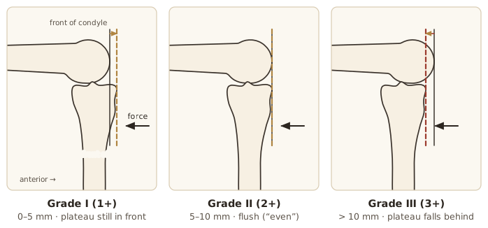
The grading figure compares the anterior aspect of the medial tibial plateau with the anterior aspect of the medial femoral condyle. Grade I: the TibiaThe shinbone, the larger bone of the lower leg.General meaning, not from the slides remains anterior to the FemurThe thighbone.General meaning, not from the slides. Grade II: the tibia is even with the femur. Grade III: the slide caption is cut off after “the tibia”; slide 17 says the plateau falls posterior. The figure labels the three positions 1+, 2+ (Even) and 3+, and shows a Force arrow with dashed lines for the three positions.
The grade limits 5 mm and 10 mm each appear in two grades on the slide (0 to 5 and 5 to 10; 5 to 10 and more than 10). The slide does not say which grade owns the boundary value.
Imaging
| Investigation | What the slides say |
|---|---|
| Plain RadiographAn X-ray picture.General meaning, not from the slides | Usually negative; they may identify a bony AvulsionA pull-off injury of the ligament at its bony insertion site. that can be reattached |
| Stress radiographsX-rays taken while a force is applied to the joint, to show how far the bones shift.General meaning, not from the slides | Compare favorably with clinical examination techniques in the diagnosis of PCLPosterior cruciate ligament: runs from the medial femoral condyle inside the notch to the back of the tibial plateau. It is the primary restraint to the tibia sliding backwards. injury |
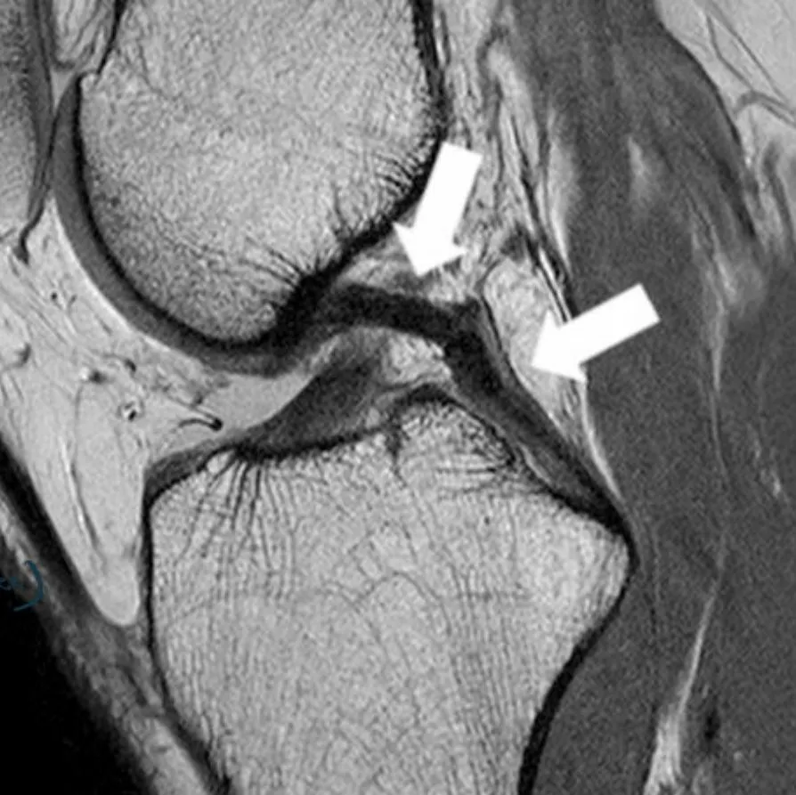
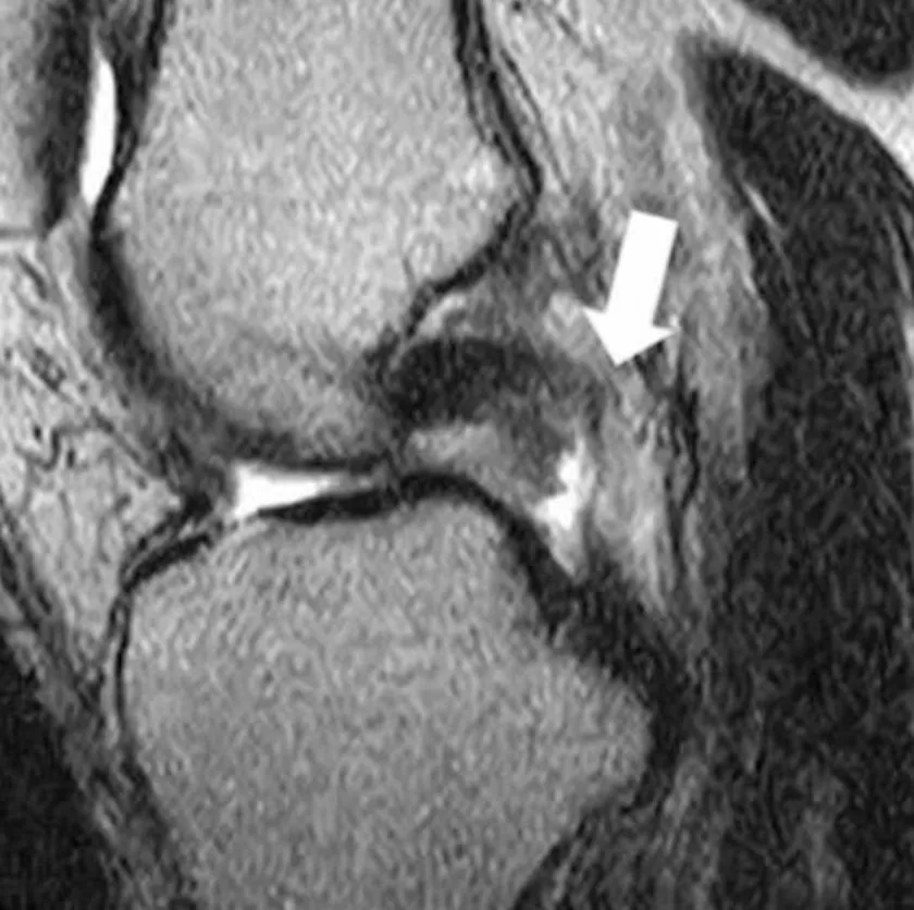
| MRIMagnetic resonance imaging: a scan that shows soft tissues such as ligaments in detail.General meaning, not from the slides | Helpful to confirm the diagnosis of a PCL rupture and to evaluate the remaining structures of the knee. Extremely SensitiveHow good a test is at picking up a problem that is really there.General meaning, not from the slides (97%) for identifying PCL tears, but not as sensitive (67%) in differentiating partial from Complete tearA tear through the whole ligament.General meaning, not from the slides |
| Bone scanAn imaging test that shows where bone is under extra stress or repair.General meaning, not from the slides | Demonstrates increased SubchondralThe bone just underneath the joint cartilage.General meaning, not from the slides stress resulting from changes in knee KinematicsThe way a joint moves.General meaning, not from the slides after PCL injury. The increased stresses may predispose the knee to early DegenerationGradual wear and breakdown of tissue, such as joint cartilage.General meaning, not from the slides, and some surgeons use the abnormal bone scan as an indication of the need for OperativeInvolving surgery.General meaning, not from the slides PCL StabilizationMaking a joint steady again, here by surgery on the PCL.General meaning, not from the slides |
MRI examples (both SagittalA view that slices the body into left and right parts, seen from the side.General meaning, not from the slides PD-weightedProton-density weighted: an MRI setting that shows ligaments, cartilage and fluid clearly.General meaning, not from the slides MR images): the normal PCL (arrow) and a complete PCL tear (arrow).
Non-surgical Care and Surgery
Frequency and non-surgical care
- PCLPosterior cruciate ligament: runs from the medial femoral condyle inside the notch to the back of the tibial plateau. It is the primary restraint to the tibia sliding backwards. injuries are reported to range between 3% and 37% of all knee LigamentA tough band of fibrous tissue that joins one bone to another and steadies a joint.General meaning, not from the slides injuries.
- Athletic injuries are most likely to result in isolated PCL injury, while motor vehicle accidents are more associated with multiple-ligament damage.
- In general, most PCL injuries are Partial tearA tear through only part of the ligament.General meaning, not from the slides and can be treated NonoperativeTreated without surgery.General meaning, not from the slides (Grade IPCL injury with 0 to 5 mm of posterior translation; the medial tibial plateau stays anterior to the medial femoral condyle. and II tears), as few will develop symptoms of Functional instabilityThe knee feeling as if it gives way during everyday or sporting activity.General meaning, not from the slides.
- Healing of the chronically injured PCL is higher when compared to ACLAnterior cruciate ligament: the knee ligament that mainly stops the tibia sliding forward on the femur.General meaning, not from the slides injuries and appears more favorable in low to mid-grade tears.
- Grade I and II tears have a rapid recovery.
| Tear | Goal: return to sport |
|---|---|
| Grade I and II | Within 2 to 4 weeks after injury |
| Grade IIIPCL injury with more than 10 mm of posterior translation; the medial tibial plateau falls posterior to the medial femoral condyle. | At about 3 months after the tear |
When is surgery indicated?
- The first line of the slide is cut off. It says that there is no clear consensus as to when ReconstructionSurgery that rebuilds a torn ligament, usually with a graft.General meaning, not from the slides is indicated for isolated PCL injuries.
| Injury | Recommendation on the slide |
|---|---|
| Acute isolated grade I or II PCL injury | The common recommendation is nonoperative rehabilitation (Rehabilitation Protocol 53.1) |
| Acute isolated grade III injury | The clear indication for surgery is an AvulsionA pull-off injury of the ligament at its bony insertion site. or “pull-off” injury of the ligament at the bony insertion site |
- Less clear are the indications for surgical treatment of mid-… (the slide is cut off here).
Link to imaging: RadiographAn X-ray picture.General meaning, not from the slides can show a bony avulsion that can be reattached, which is the clear surgical case for an acute isolated grade III injury.
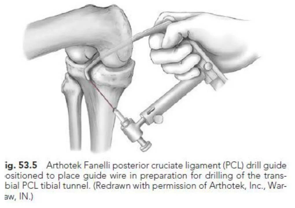
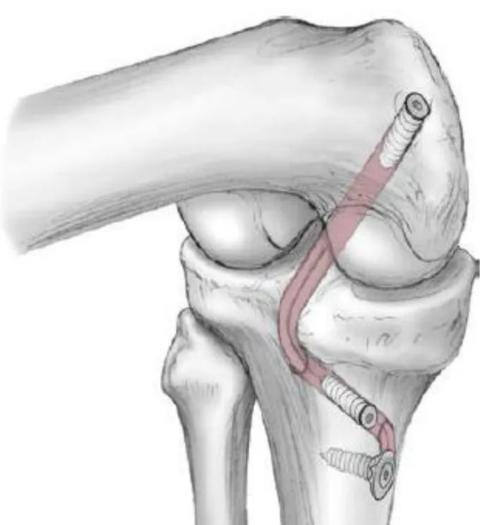
Slide 25 shows two surgical drawings: Fig. 53.5, an Arthotek Fanelli PCL Drill guideAn instrument that aims the drill so the bone tunnel is made in the right place.General meaning, not from the slides positioned to place the Guide wireA thin wire placed first to mark the path the drill will follow.General meaning, not from the slides in preparation for drilling of the Trans-tibialPassing through the tibia (shinbone).General meaning, not from the slides PCL Tibial tunnelA channel drilled in the shinbone for the new ligament (graft) to pass through.General meaning, not from the slides, and a second, uncaptioned drawing of a reconstructed ligament.
PCL Rehabilitation
Exercise stress on the PCL
- Stress on the PCLPosterior cruciate ligament: runs from the medial femoral condyle inside the notch to the back of the tibial plateau. It is the primary restraint to the tibia sliding backwards. can occur during MidrangeThe middle part of a joint's movement, away from the extremes.General meaning, not from the slides Weight bearingPutting body weight through the leg. Non weight-bearing means no body weight goes through it.General meaning, not from the slides ResistedDone against an added force such as a weight, band or the therapist's hand.General meaning, not from the slides knee ExtensionStraightening a joint, for example straightening the knee.General meaning, not from the slides.
- Wilk reported a Shear forceA force that makes one surface slide across another, here the tibia sliding backward on the femur.General meaning, not from the slides during resisted knee extension between 100° and 40°, with the peak at 85° to 95°.
- The PCL shear forces from 60° to 0° were lower, and knee extension is performed most safely in this range.
- Resisted knee FlexionBending a joint, for example bending the knee.General meaning, not from the slides exercises should be approached with caution until adequate healing of the PCL (at least 4 months after PCL ReconstructionSurgery that rebuilds a torn ligament, usually with a graft.General meaning, not from the slides), because of the excessively high PCL stress that occurs with HamstringsThe muscle group on the back of the thigh that bends the knee.General meaning, not from the slides contractions.
General guidelines
(Slides 27 and 28 carry the same list.)
- No Open chainOpen kinetic chain exercise: the foot is free to move, as in a seated leg extension.General meaning, not from the slides.
- Caution against TranslationStraight sliding of one bone on another, for example the tibia sliding backward (posterior) or forward (anterior) on the femur.General meaning, not from the slides (by gravity, muscle action).
- No Continuous passive motionA machine that moves the joint over and over without effort from the patient.General meaning, not from the slides.
- Resistance for hip PREProgressive resistance exercise: strengthening work where the load is increased step by step.General meaning, not from the slides (PREs) is placed above the knee for hip AbductionMoving a limb away from the midline of the body.General meaning, not from the slides and AdductionMoving a limb toward the midline of the body.General meaning, not from the slides; resistance may be DistalFarther from the trunk or the point of attachment of a limb.General meaning, not from the slides for hip flexion.
Phase 1: weeks 0–4
Goals:
- Protect healing bony and soft tissue structures.
- Minimize the effects of ImmobilizationKeeping a joint still, for example in a brace or cast.General meaning, not from the slides.
- Early protected ROMRange of motion: how far a joint can move.General meaning, not from the slides (protection against Posterior sagThe tibia dropping back relative to the femur, for example under gravity when the knee is bent.General meaning, not from the slides).
- PREs for QuadricepsThe large muscle group on the front of the thigh that straightens the knee.General meaning, not from the slides, hip and calf, with emphasis on limiting Patellofemoral jointThe joint between the kneecap and the thighbone.General meaning, not from the slides compression and posterior tibial translation.
- Patient education for a clear understanding of the limitations and expectations of the rehabilitation process, and the need for supporting the Proximal tibiaThe upper end of the shinbone, just below the knee.General meaning, not from the slides and avoiding sag.
Bracing:
- Brace locked at 0 degrees for 1 week.
- At 1 week after surgery, the brace is unlocked for Passive ROMJoint movement done by someone else (or by the patient's other hand) while the muscles stay relaxed.General meaning, not from the slides done by the physical therapist or athletic trainer.
- The patient is instructed in self-administered passive ROM with the brace on, with emphasis on supporting the proximal tibia.
Weight bearing: as tolerated with crutches, brace locked in extension.
Special considerations: a pillow under the ProximalCloser to the trunk or the point of attachment of a limb.General meaning, not from the slides PosteriorToward the back of the body.General meaning, not from the slides TibiaThe shinbone, the larger bone of the lower leg.General meaning, not from the slides at rest to prevent posterior sag.
Therapeutic exercises:
- Patellar mobilizationGentle hands-on gliding of the kneecap to keep it moving freely.General meaning, not from the slides
- ProneLying face down.General meaning, not from the slides passive flexion and extension
- Quadriceps setTightening the thigh muscle with the knee straight, pressing the back of the knee down.General meaning, not from the slides
- SLRStraight leg raise: lifting the straight leg off the bed, which needs a good quadriceps contraction.General meaning, not from the slides (SLR)
- Hip abduction and adduction
- Ankle pumpsMoving the foot up and down at the ankle to keep the circulation and movement going.General meaning, not from the slides
- Hamstring and calf stretching
- Calf exercise with TherabandA brand of stretchy resistance band used for strengthening.General meaning, not from the slides, progressing to Standing calf raiseRising up onto the toes while standing, to strengthen the calf muscles.General meaning, not from the slides with full knee extension
- Standing Hip extensionMoving the thigh backward at the hip.General meaning, not from the slides from neutral
- Functional electrical stimulationElectrical stimulation that makes a muscle contract so it can be used in a movement.General meaning, not from the slides (may be used for trace to poor quadriceps contraction)
Phase 2: weeks 4–12
Criteria for progression to Phase 2:
- Good quadriceps control (good quadriceps set, no sag with SLR).
- Approximately 60 degrees of knee flexion.
- Full knee extension.
- No signs of active InflammationThe body's response to injury: swelling, warmth, redness and pain.General meaning, not from the slides.
Goals:
- Increase ROM (flexion).
- Restore normal GaitThe way a person walks.General meaning, not from the slides.
- Continue quadriceps strengthening and hamstring flexibility.
PCL rehabilitation protocol by week
| Time | Visits (slide wording) | Treatment | Milestones |
|---|---|---|---|
| Week 1 | Total visits: 1 | NMESNeuromuscular electrical stimulation: electrical current through skin electrodes that makes a muscle contract.General meaning, not from the slides (see guidelines) · Quad sets · SLR · Patellar mobilization · HEPHome exercise programme: the exercises the patient does at home.General meaning, not from the slides: patellar mobilization 30–50X, quad sets and SLR 3x10 (3x per day) | Good quadriceps contraction · Superior patellar glideThe kneecap sliding upward, toward the head.General meaning, not from the slides · AmbulatingWalking.General meaning, not from the slides PWBPartial weight bearing: only part of the body weight is put through the leg.General meaning, not from the slides with crutches with OrthosisA brace that supports a joint. A postop orthosis is the brace worn after surgery.General meaning, not from the slides locked |
| Week 2 | 2–3 visits/week; total visits: 3–4 | Portal/incision mobilizationGentle hands-on movement of the tissue around the surgical keyhole or cut to keep it from stiffening.General meaning, not from the slides as needed · SAQShort arc quad: straightening the knee through a short range, here 30° to 0°.General meaning, not from the slides 30°–0° | Full extension · Flexion to 60 · SLR without Quadriceps lagThe knee cannot be held fully straight on its own during a straight leg raise because the quadriceps is not contracting fully.General meaning, not from the slides (full quadriceps contraction) |
| Weeks 3–5 | 2–3 visits/week; total visits: 9–13 | Prone knee flexion, therapist assisted 0–60° · SupineLying face up.General meaning, not from the slides knee flexion holding tibia forward · OKC 60–0° · Stationary bike for ROM, easy · Gait training PWB with crutches, no orthosis | Flexion to 110° · Quad strength >60% of Involved and uninvolvedInvolved is the injured or operated leg; uninvolved is the other leg, used for comparison.General meaning, not from the slides · Wean from orthosis, normalize gait crutches (slide wording) |
| Weeks 6–10 | 2–3 visits/week; total visits: 19–28 | Stationary bike, easy · Begin Closed chainClosed kinetic chain exercise: the foot is fixed on the ground or a surface, as in a squat.General meaning, not from the slides if good quad control: Wall sits and wall squatsClosed chain exercises with the back against a wall: holding a bent-knee position (sit) or sliding up and down (squat).General meaning, not from the slides, wall squats 0°–45° | Normal gait without crutches · Quad strength >80% of uninvolved |
| Week 12 | Twice per week to rechecks (slide wording) | Progress exercise intensity and duration · 0°–90° hamstring exercises against gravity | Pain-free AROMActive range of motion: how far a joint moves using the patient's own muscle power.General meaning, not from the slides to within 10 of uninvolved · Maintaining or increasing quadriceps strength (>= 90%) |
| Week 16 | Twice per week to rechecks (slide wording) | Begin running progression with Functional braceA brace that supports the knee during activity, here during the running progression.General meaning, not from the slides (see note) · PRE-hamstring curls 0°–90° · Transfer to fitness facility (if all milestones are met) | Full ROM (compared to uninvolved) · Maintaining quadriceps strength ≥ 95% |
| Week 20 | Rechecks; total visits: 25–44 | Return to sport transition · ProprioceptiveTo do with the sense of where a joint is in space without looking.General meaning, not from the slides, Static balanceStaying balanced while standing still.General meaning, not from the slides, Dynamic balanceStaying balanced while moving.General meaning, not from the slides, functional activities: slow to fast speed, low to high force, controlled to uncontrolled | Global reportThe patient's overall self-reported rating of how the knee is doing, shown as a percentage.General meaning, not from the slides >70% · KOS ADLSKnee Outcome Survey, Activities of Daily Living Scale: a questionnaire on how well the knee copes with everyday tasks.General meaning, not from the slides >90% |
Note on the slide: MVICMaximum Volitional Isometric Contraction: the patient extends the involved leg as hard as possible with the knee held isometrically at 30° flexion. = Maximum Volitional IsometricA muscle contraction in which the muscle tenses but the joint does not move.General meaning, not from the slides Contraction. The patient is asked to volitionally extend the involved leg as hard as possible while the knee is held isometrically at 30° knee flexion. Side-to-side comparison: (involved / uninvolved) × 100 = % MVC (the slide writes “% MVC” here).
How to read the weeks: early weeks are about quad control and ROM (full extension and flexion to 60 by week 2, flexion to 110° by weeks 3–5). Closed chain is listed from weeks 6–10 “if good quad control”, and the running progression at week 16.
Open chain is mentioned twice. The general guidelines say “No open chain exercises”, yet the weeks 3–5 row lists “OKC 60–0°”, and the exercise-stress slide calls knee extension safest from 60° to 0°. The slides do not explain how these fit together, so learn both.
Bracing and weight bearing, weeks 4–8
Bracing:
- 4–6 weeks: the brace is unlocked for controlled gait training only (the patient may walk with the brace unlocked while attending physical therapy or when at home).
- 6–8 weeks: the brace is unlocked for all activities.
- 8 weeks: the brace is discontinued (as allowed by the physician).
Weight bearing:
- 4–8 weeks: weight bearing as tolerated with crutches.
- 8 weeks: may discontinue crutches if the patient exhibits no quadriceps lag with SLR.
Also listed on the slide (no heading, after the weight-bearing items):
- Full knee extension
- Knee flexion 90–100 degrees
- Normal gait pattern (the patient can use one crutch or cane until normal gait is achieved)
Two bracing timelines. Phase 1 says the brace is locked at 0 degrees for 1 week, then unlocked for passive ROM with the therapist. The weeks 4–8 slide has the brace unlocked for gait training at 4–6 weeks and discontinued at 8 weeks. The weeks 3–5 row of the protocol table says gait training “no orthosis” and a milestone to “wean from orthosis”. The slides do not reconcile these, so keep each time frame with its own slide. Weight bearing is also worded differently: “PWB with crutches” in the protocol table versus “as tolerated with crutches” in the guidelines.
NMES guideline
- Electrodes placed over the proximal LateralAway from the middle of the body, the outer side.General meaning, not from the slides quadriceps and distal MedialToward the middle of the body, the inner side.General meaning, not from the slides quadriceps. (Modify distal electrode placement by not covering the SuperiorAbove, toward the head.General meaning, not from the slides medial (VMOVastus medialis obliquus: the part of the quadriceps on the inner side just above the kneecap.General meaning, not from the slides) Arthroscopy portalA small keyhole incision through which instruments and a camera enter the joint during arthroscopic surgery.General meaning, not from the slides until stitches are removed and skin is healed.)
- Stimulation parameters: 2500 HzHertz: cycles per second, the frequency of the stimulation current.General meaning, not from the slides, 75 BurstsShort packets of electrical pulses delivered together.General meaning, not from the slides, 2 sec RampThe gradual build-up of the current at the start of each contraction.General meaning, not from the slides, 12 sec on, 50 sec rest, intensity to max tolerable (at least 50% MVIC), 10 contractions per session. 3 sessions per week until quadriceps strength MVIC is 80% of uninvolved.
- Stimulation performed isometrically at 30° (dependent on GraftTissue used to replace or rebuild a ligament.General meaning, not from the slides site).
Criteria for return to sports
- Full, pain-free ROM
- Satisfactory clinical examination
- Quadriceps strength 85% of the ContralateralOn the opposite side of the body; here the other, uninjured leg.General meaning, not from the slides leg
- Functional testingTests that check how well the leg performs real-life movements, compared with the other leg.General meaning, not from the slides 85% of the contralateral leg
- No change in LaxityLooseness: extra give in a joint or ligament.General meaning, not from the slides testing
Quadriceps strength numbers at a glance
| Where on the slides | Quadriceps strength figure |
|---|---|
| Weeks 3–5 milestone | >60% of uninvolved |
| Weeks 6–10 milestone | >80% of uninvolved |
| NMES: continue until | 80% of uninvolved (MVIC) |
| Week 12 milestone | >= 90% (maintaining or increasing) |
| Week 16 milestone | ≥ 95% (maintaining) |
| Criteria for return to sports | 85% of the contralateral leg |
The slides do not explain how the 85% return-to-sports criterion relates to the ≥ 90% and ≥ 95% milestones, so learn which number belongs to which time point.