Rehabilitation of Hip Injuries in Sport
Quiz this lecture (157 questions)This lecture covers the rehabilitation of hip injuries in sport. It starts with the clinical examination of the hip (history, age and gender, pain, aggravating and relieving factors, tests and measures), then goes through the main conditions: buttock pain (Hamstring syndromePain from sciatic nerve entrapment near the ischial tuberosity, where fibrous bands from the biceps insertion can form a tissue envelope around the nerve., Piriformis syndromeButtock pain from compression and irritation of the sciatic nerve as it courses out from under the piriformis muscle.), StrainAn overstretch or tear of a muscle or its tendon. Graded I to III, where grade III is a complete tear.General meaning, not from the slides with its three-phase rehabilitation and return-to-sport criteria, Gluteal tendinopathyTendon problem of the gluteus medius and minimus at the greater trochanter, causing lateral hip pain. The slide title also calls it greater trochanter pain syndrome. and its rehabilitation program, groin pain (Adductor tendinopathyPain and degeneration of the tendons of the inner thigh (adductor) muscles, usually felt in the groin.General meaning, not from the slides, Osteitis pubisInflammation of the pubic symphysis with bone changes around it, giving groin and pubic pain that is frequently severe., Athletic pubalgiaAlso called sport's hernia or hockey hernia: weakening or tearing of the abdominal wall structures, creating an inside-out hernia in the dorsal wall of the inguinal canal.), Labral tearA tear in the labrum of the hip. The slides classify tears as traumatic vertical or degenerative horizontal.General meaning, not from the slides and Femoroacetabular impingementAn abnormal shape of the proximal femur and acetabulum that raises contact forces in hip flexion, potentially injuring the labrum and cartilage and causing pain. (FAI), and Avulsion fractureFailure of the bone at the tendinous insertion rather than the tendon itself, so a piece of bone is pulled off.. It ends with the general rehabilitation toolkit: hip mobility and flexibility, the Powers ProgramThe slides' program of eight phases for gluteus maximus activation, strengthening and function, with an emphasis on triplanar GMax function. for Gluteus maximusThe largest buttock muscle, which extends the hip and rotates it outward.General meaning, not from the slides activation and strengthening, and advanced functional training.
Clinical examination of the hip
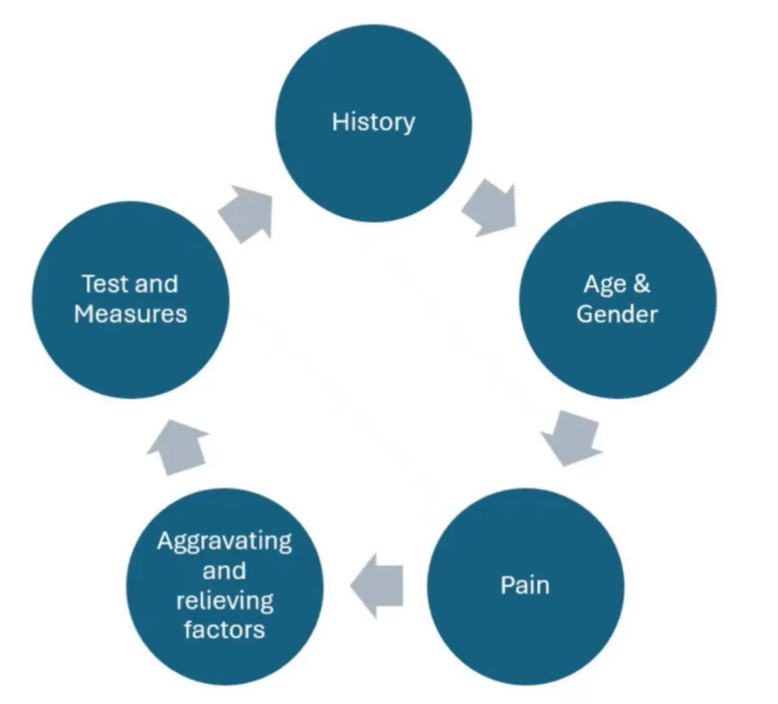
The examination is a cycle of five steps: History → Age and gender → Pain → Aggravating and relieving factors → Tests and measures.
History
- The Differential diagnosisSorting through the possible causes of a symptom to work out which one fits best.General meaning, not from the slides of hip pain includes IntraarticularInside the joint.General meaning, not from the slides and ExtraarticularOutside the joint, in the tissues around it.General meaning, not from the slides pathologies.
- In addition, other disorders unrelated to the hip can mimic hip pathology.
Age and gender
| Age / group | Condition |
|---|---|
| 15–25 years | Osteochondritis dissecansA condition in which a piece of joint cartilage and the bone beneath it loosens from the joint surface.General meaning, not from the slides |
| 18–40 years | Labral tearA tear in the labrum of the hip. The slides classify tears as traumatic vertical or degenerative horizontal.General meaning, not from the slides |
| Over 40 years, especially males | Labral cystA fluid-filled sac in or next to the labrum.General meaning, not from the slides, SacrumThe triangular bone at the base of the spine, wedged between the two hip bones.General meaning, not from the slides pathologies, and Stress fractureA small crack in a bone caused by repeated loading rather than one single injury.General meaning, not from the slides of the femur or pelvis |
| Young athletic females | Greater risk of stress fractures in the ProximalCloser to the trunk or to the point where a limb or structure attaches.General meaning, not from the slides femoral region, associated with the Female triadAs listed on the slide: eating disorders, dysmenorrhea and osteoporosis. It is linked with stress fractures of the proximal femoral region in young athletic females. (eating disorders, DysmenorrheaPainful menstrual periods.General meaning, not from the slides and OsteoporosisA condition in which bones become thin and weak.General meaning, not from the slides) |
Pain
- There is a high incidence of Referred painPain felt in one place that actually comes from a problem somewhere else.General meaning, not from the slides both to and from the hip region. The location or type of pain may or may not be helpful in all diagnoses to identify the type or presence of hip pathology.
Quality of pain
| Quality | Associated with |
|---|---|
| Aching | BursitisInflammation of a bursa.General meaning, not from the slides, TendinopathyPain and degeneration of a tendon, usually from overload.General meaning, not from the slides, ArthritisInflammation of a joint, causing pain and stiffness.General meaning, not from the slides |
| Sharp | Labral tears or Loose bodiesSmall fragments of bone or cartilage floating free inside a joint.General meaning, not from the slides |
| Burning | Nerve entrapmentA nerve that is squeezed or trapped by the tissues around it.General meaning, not from the slides; can be accompanied by ParesthesiasAbnormal skin sensations such as tingling or pins and needles.General meaning, not from the slides, numbness, and/or weakness, sweating and hair loss in sympathetic involvement |
Site of pain
| Site | Can come from |
|---|---|
| Groin pain | CFJCoxafemoral joint: the hip joint, where the head of the femur sits in the socket of the pelvis.General meaning, not from the slides and LabrumThe ring of cartilage around the rim of the hip socket (acetabulum). It deepens the socket and helps seal the joint.General meaning, not from the slides injury, Symphysis pubisThe joint at the front of the pelvis where the two pubic bones meet.General meaning, not from the slides lesions, Adductor tendinopathyPain and degeneration of the tendons of the inner thigh (adductor) muscles, usually felt in the groin.General meaning, not from the slides, Iliopectineal bursitisInflammation of the iliopectineal bursa, a cushion at the front of the hip next to the iliopsoas tendon.General meaning, not from the slides, incompetent abdominal wall, or any number of urological, gynecological, neurovascular or organic lesions of the abdominal cavity and pelvic region, LumbarRelating to the lower back, the part of the spine between the chest and the pelvis.General meaning, not from the slides and/or SIJSacroiliac joint: the joint between the sacrum and the hip bone (ilium) on each side of the pelvis.General meaning, not from the slides |
| Buttock pain | SIJ dysfunction, Gluteal bursitisInflammation of a bursa beneath the gluteal (buttock) muscles.General meaning, not from the slides, HamstringsThe muscle group at the back of the thigh that bends the knee and extends the hip.General meaning, not from the slides tendinopathy, Hamstring syndromePain from sciatic nerve entrapment near the ischial tuberosity, where fibrous bands from the biceps insertion can form a tissue envelope around the nerve., and/or SciaticaPain that runs along the path of the sciatic nerve, from the buttock down the back of the leg.General meaning, not from the slides |
| Posterolateral hip pain (Greater trochanterThe bony point on the outer side of the upper femur, where the gluteal muscles attach.General meaning, not from the slides) | Trochanteric bursitisInflammation of the bursa over the greater trochanter, the bony point on the outer side of the hip.General meaning, not from the slides, Gluteal tendinopathyTendon problem of the gluteus medius and minimus at the greater trochanter, causing lateral hip pain. The slide title also calls it greater trochanter pain syndrome., lumbar spine |
Aggravating and relieving factors
- Increased groin pain with coughing, sneezing or straining can suggest a HerniaA bulge of tissue pushing through a weak spot in the wall that normally holds it in.General meaning, not from the slides, or tendinopathy of the Adductor longusA thigh muscle on the inner side that pulls the leg toward the midline. The slide groups it with the adductor brevis.General meaning, not from the slides or Rectus abdominisThe straight muscle at the front of the abdomen that flexes the trunk.General meaning, not from the slides.
- Provocation of the buttock pain suggests involvement of the Nerve rootThe part of a spinal nerve that leaves the spine. Lumbar and sacral roots supply the lower back, pelvis and legs.General meaning, not from the slides in a Prolapsed diskA spinal disk whose soft center has pushed out and can press on a nerve root.General meaning, not from the slides.
- If pain is only experienced at night, always rule out systemic disease and/or tumor, such as an Osteoid osteomaA small, benign (non-cancerous) bone tumor.General meaning, not from the slides.
- If experienced during the day, consider patient position:
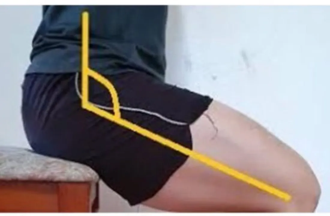
- Groin pain increased by sitting could be associated with a lesion of the anterosuperior acetabular labrum.
- Buttock pain increased with sitting could be related to Ischial bursitisInflammation of the bursa over the ischial tuberosity, the bone you sit on.General meaning, not from the slides, hamstring syndrome, gluteal bursitis, or a lumbar disk pathology.
Position hook: sitting + groin = labrum; sitting + buttock = ischial bursitis / hamstring syndrome / gluteal bursitis / lumbar disk.
Tests and measures
- The hip examination should include screening of the lumbar spine and SIJs, and swelling in the CFJ.
- Gluteal muscle AtrophyShrinking and wasting of a muscle.General meaning, not from the slides suggests S1 and/or S2 root levels.
- Nodular formations over the sacrum suggest benign LipomaA benign (non-cancerous) lump of fat tissue.General meaning, not from the slides.
- Lumbar movement screening (lumbosacral roots).
- The SLRDIStraight leg raise, distal initiation: the straight leg raise test with ankle and foot dorsiflexion and the head and neck in neutral, so the test starts at the far end. (SLRDI).
- The SDISlump distal initiation: the slump test started from the far end (distally), used to provoke nerve symptoms.General meaning, not from the slides (SDI).
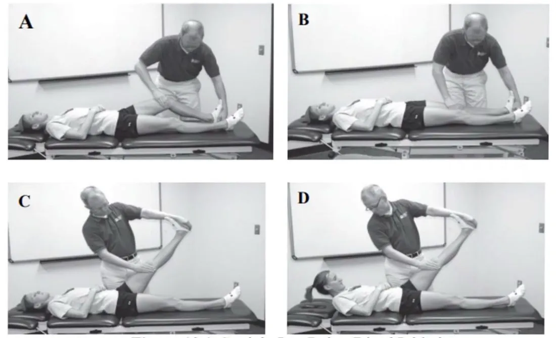
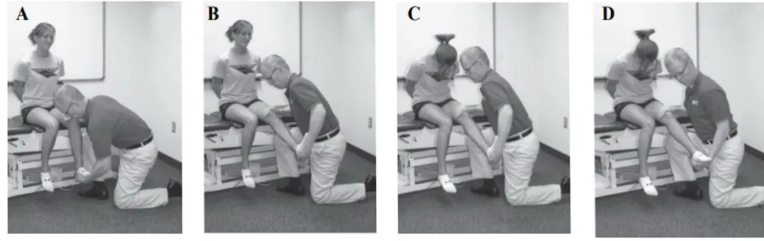
Other tests shown on the slides (photos only, no text):
- The ASLRActive straight leg raise: the patient lifts the straight leg. The slide labels this test as a sacroiliac joint test. (ASLR) test, labelled SIJ (slide 10).
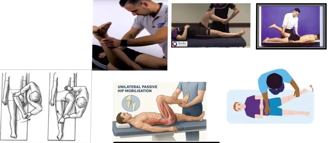
- Hip examination, passive movements (slide 11). One picture is labelled “unilateral passive hip mobilisation”.
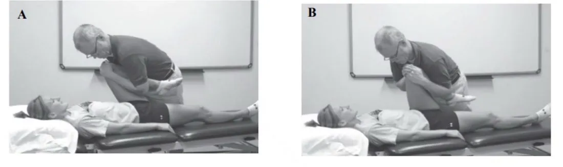
- The Modified circumduction testA hip test labelled on the slides as a hip impingement test. It is used as a provocative maneuver for labral tears., labelled hip impingement (slide 12).
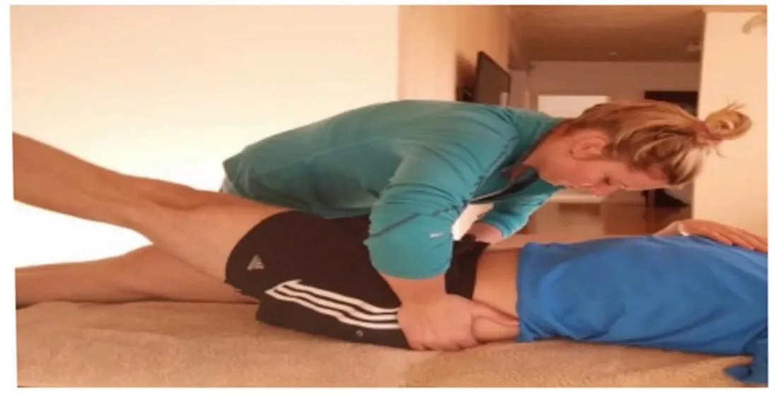
Resisted movements
| Position | Movement (muscle) |
|---|---|
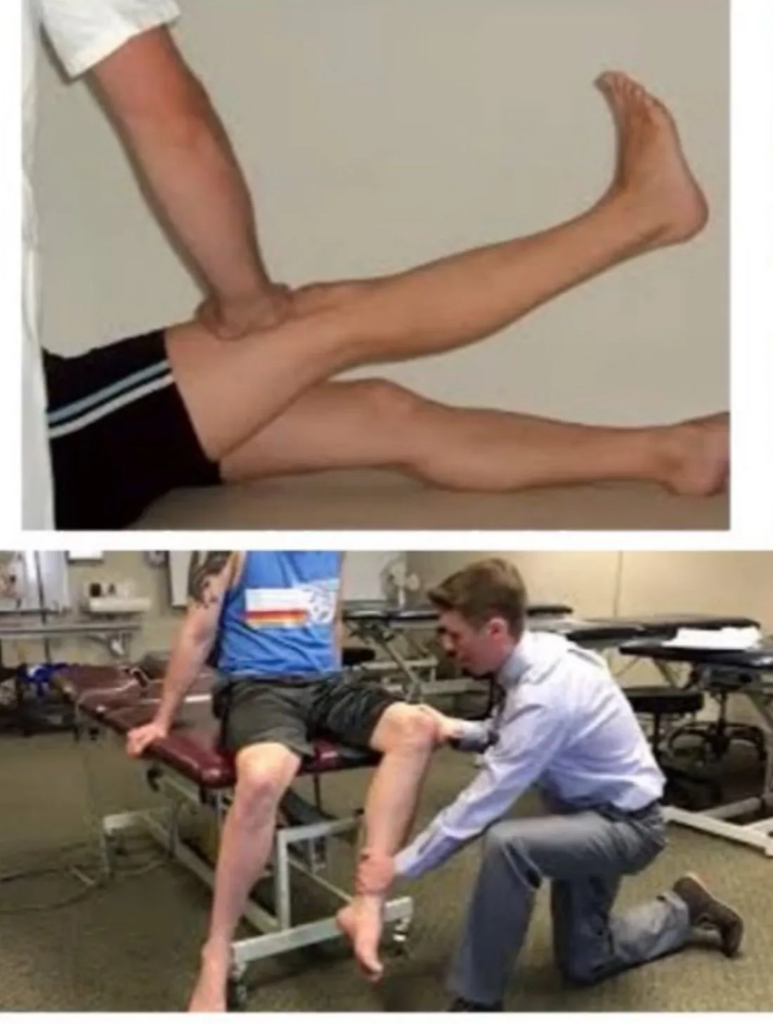
| Supine | a. Hip flexion (PsoasA deep muscle from the lower spine to the top of the femur that flexes the hip.General meaning, not from the slides) · b. Straight leg raiseLifting the straightened leg, either as a test (passive or active) or as a muscle test of the hip flexors.General meaning, not from the slides (Rectus femorisThe muscle at the front of the thigh that flexes the hip and extends the knee.General meaning, not from the slides) · c. External rotation (in 90° flexion) · d. Internal rotation (in 90° flexion) · e. Double-leg BridgeLifting the hips up from lying on the back, or holding a body-bridge position, to work the buttocks, hamstrings and trunk.General meaning, not from the slides · f. Single-leg bridge |
| Side-lying | Hip abduction in neutral (TFLTensor fasciae latae: a small hip muscle on the outer upper thigh that tenses the iliotibial band.General meaning, not from the slides / Gluteus mediusThe gluteal muscle on the outer side of the hip that abducts the hip and steadies the pelvis.General meaning, not from the slides) |
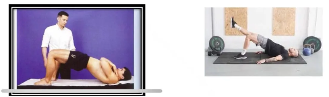
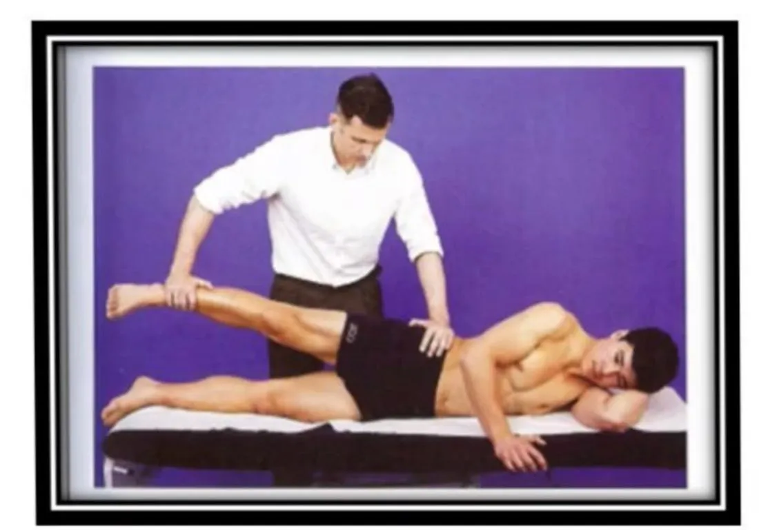
Buttock pain
Slide 15 lists the sources of buttock pain: Sciatic nerveThe largest nerve in the body. It runs from the lower back through the buttock and down the back of the leg.General meaning, not from the slides, Piriformis syndromeButtock pain from compression and irritation of the sciatic nerve as it courses out from under the piriformis muscle., Hamstring syndromePain from sciatic nerve entrapment near the ischial tuberosity, where fibrous bands from the biceps insertion can form a tissue envelope around the nerve., Ischial bursitisInflammation of the bursa over the ischial tuberosity, the bone you sit on.General meaning, not from the slides, CFJCoxafemoral joint: the hip joint, where the head of the femur sits in the socket of the pelvis.General meaning, not from the slides, LabrumThe ring of cartilage around the rim of the hip socket (acetabulum). It deepens the socket and helps seal the joint.General meaning, not from the slides affliction, trochanteric / Gluteal bursitisInflammation of a bursa beneath the gluteal (buttock) muscles.General meaning, not from the slides, LumbarRelating to the lower back, the part of the spine between the chest and the pelvis.General meaning, not from the slides disk RadiculopathyPain, numbness or weakness caused by a pinched or irritated spinal nerve root.General meaning, not from the slides, SIJSacroiliac joint: the joint between the sacrum and the hip bone (ilium) on each side of the pelvis.General meaning, not from the slides, sacrococcygeal joint.
Hamstring syndrome
Cause and onset
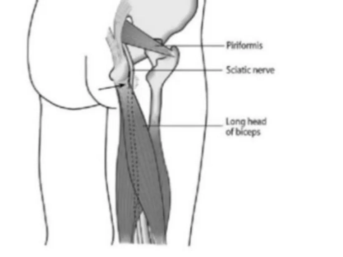
- Fibrous bands can project from the biceps bony InsertionThe place where a tendon attaches to a bone.General meaning, not from the slides, potentially creating a tissue envelope that can surround the sciatic nerve as it courses lateral to the Ischial tuberosityThe bony point at the bottom of the pelvis that you sit on. The hamstrings attach here.General meaning, not from the slides, leading to hamstring syndrome.
- Hamstring syndrome develops as the result of sciatic nerve entrapment.
- It is frequently preceded by episodic HamstringsThe muscle group at the back of the thigh that bends the knee and extends the hip.General meaning, not from the slides injury and/or a history of low back pain or surgery that may predispose the sciatic nerve to a greater vulnerability for injury.
- Seen frequently in distance runners, sprinters and jumping athletes.
- The pain begins gradually and worsens after persistent physical activity.
Clinical findings
- Increased pain with sitting.
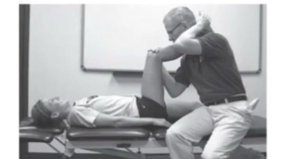
- Resisted knee flexion with the hip flexed to 90° and the knee extended to the limit is painful. The provocation may be worsened during the same test with ankle and foot DorsiflexionPulling the foot up toward the shin.General meaning, not from the slides.
- Resisted knee flexion with the hip extended (prone) is pain free.
- Local PalpationExamining by feeling with the hands.General meaning, not from the slides over the lateral ischial tuberosity is painful.
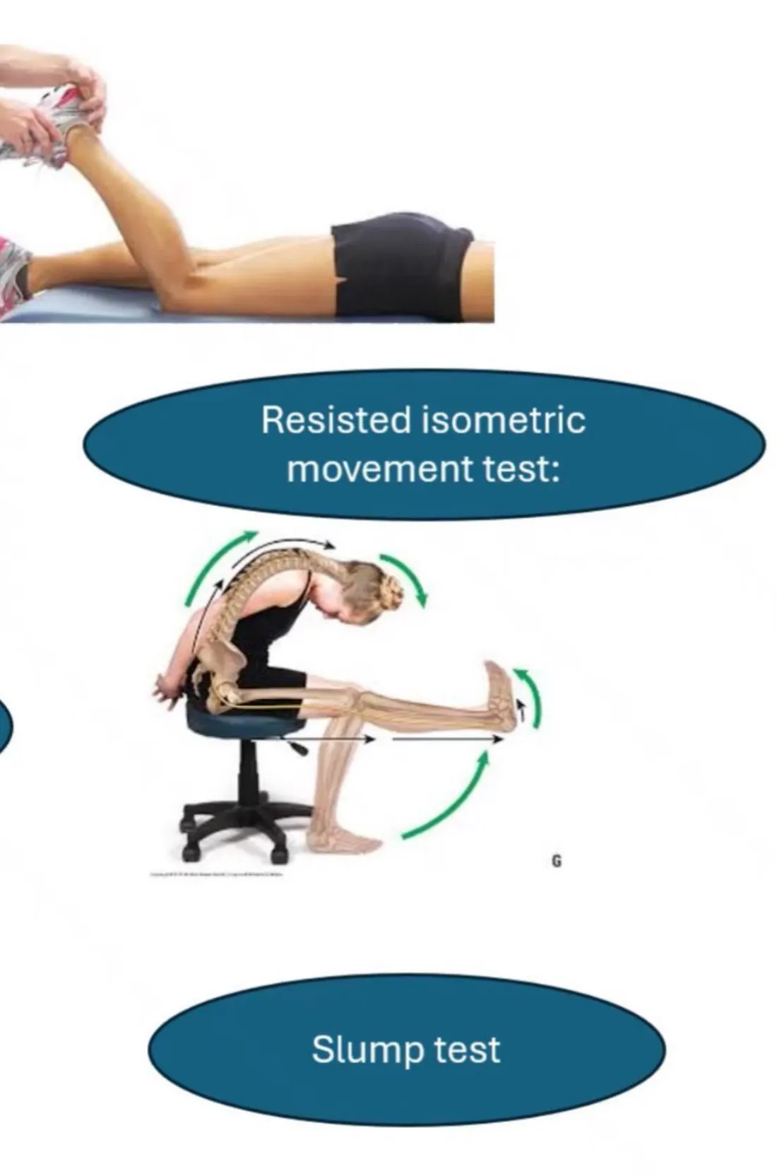
- The Straight leg raiseLifting the straightened leg, either as a test (passive or active) or as a muscle test of the hip flexors.General meaning, not from the slides and/or Slump testA seated test that stretches the nerves along the back and legs, used to see whether neural tissue reproduces symptoms.General meaning, not from the slides could be provocative, increased with ankle and foot dorsiflexion and the head and neck in neutral (DistalFarther from the trunk or from the point where a limb or structure attaches.General meaning, not from the slides initiated).
- Stretching exercises do not alleviate symptoms, frequently making them worse.
Management
- No hamstring stretching activities.
- Educate the patient to sit on a wedge with the thicker side of the wedge positioned dorsally.
- Gentle Neural mobilizationGentle movements that slide and stretch a nerve through the tissues around it.General meaning, not from the slides/manipulation of the lower extremity, distally initiated at the knee or ankle and foot.
- IontophoresisUsing a mild electrical current to drive a medication through the skin.General meaning, not from the slides applied to the gluteal region just lateral to the ischial tuberosity, with the patient in side-lying on the non-painful side and the hips and knees flexed to 90°.
- Examination and retraining of the Pelvic floor musclesThe group of muscles that form a sling across the bottom of the pelvis.General meaning, not from the slides in females with hamstring syndrome, stating “The concept of Regional interdependenceThe slide's concept that pain is somehow related to a more remote dysfunction. is highlighted in which pain is somehow related to a more remote dysfunction”.
- Severe cases may require open surgical release.
Piriformis syndrome (Deep Gluteal Pain Syndrome)
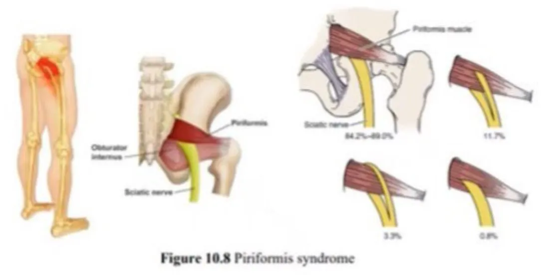
Cause
- Can produce buttock pain as a consequence of sciatic nerve compression and irritation.
- This can occur as the nerve courses caudally out from under the inferior edge of the PiriformisA small muscle deep in the buttock. The sciatic nerve runs beside it.General meaning, not from the slides muscle.
- Nerve irritation can begin with blunt trauma to the buttock region and/or overuse in activities that place the sciatic nerve under tension, such as running.
- 14% of all individuals exhibit a perforation of the piriformis muscle by the peroneal branch of the sciatic nerve, which causes nerve compression each time the piriformis is used.
- Persistent sciatic arteries or venous AneurysmA bulge in the wall of a blood vessel.General meaning, not from the slides can compress the nerve in the same location.
The figure on slide 19 shows four nerve–piriformis relationships labelled 84.2%–89.0%, 11.7%, 3.3% and 0.8%. The slide text itself gives only the 14% figure for perforation, and does not explain the figure’s percentages.
Clinical findings
- Increased pain in the buttock with walking activities, while sitting may decrease the pain except if sitting on hard surfaces for a long time.
- The pain can refer into the posterior thigh to the knee when more severe.
- A compressed Inferior gluteal nerveA nerve in the buttock that supplies the gluteus maximus.General meaning, not from the slides may cause Gluteus maximusThe largest buttock muscle, which extends the hip and rotates it outward.General meaning, not from the slides AtrophyShrinking and wasting of a muscle.General meaning, not from the slides.
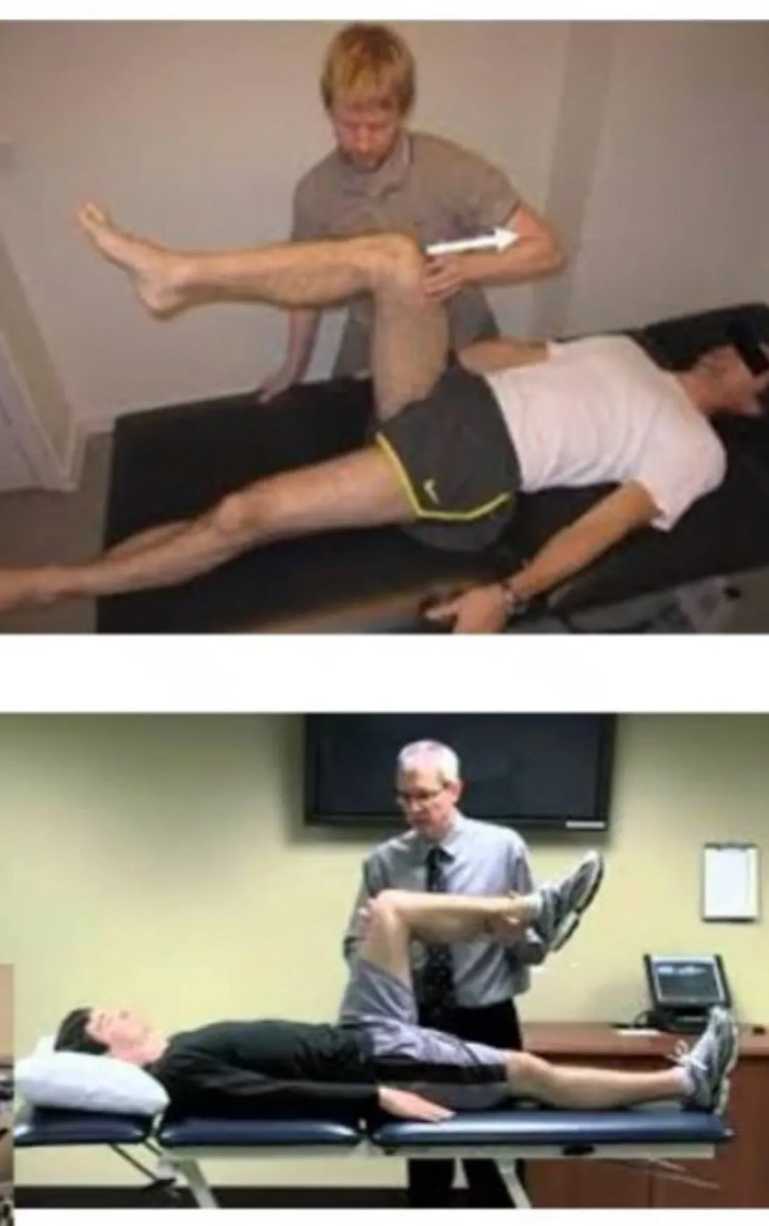
- EpineuralRelating to the outer sheath that surrounds a nerve.General meaning, not from the slides irritation and subsequent pain is produced when the hip is positioned in < 90° of flexion, abduction and internal rotation (FAIR testHip flexion, abduction and internal rotation: the position the slide uses to provoke epineural irritation in piriformis syndrome.).
- SLRDIStraight leg raise, distal initiation: the straight leg raise test with ankle and foot dorsiflexion and the head and neck in neutral, so the test starts at the far end. or SDISlump distal initiation: the slump test started from the far end (distally), used to provoke nerve symptoms.General meaning, not from the slides testing could be provocative.
FAIR (slide 20) lists flexion, abduction and internal rotation. The FADIRFlexion, adduction and internal rotation: the hip position whose pain reproduction the slide lists as a finding in femoroacetabular impingement. test used for Femoroacetabular impingementAn abnormal shape of the proximal femur and acetabulum that raises contact forces in hip flexion, potentially injuring the labrum and cartilage and causing pain. (slide 61) is flexion, adduction and internal rotation. They are two different tests.
Management
- As before, the patient is educated to avoid stretching and sitting on hard surfaces.
- Gentle neural mobilization/manipulation.
- Injection of CorticosteroidsStrong anti-inflammatory medicines, here given by injection.General meaning, not from the slides and AnalgesicsPain-relieving medicines, or the state of pain relief.General meaning, not from the slides under FluoroscopyLive X-ray imaging, used here to guide a needle to the right place.General meaning, not from the slides guidance may reduce pain and piriformis muscle activity.
- Botulinic toxinA toxin injected in tiny doses to relax a muscle.General meaning, not from the slides injection can be suggested.
- Surgical release might be required in extreme cases.
Hamstring strain
Overview and risk factors
- Common in sprinting sports as well as dancers.
- Grades of StrainAn overstretch or tear of a muscle or its tendon. Graded I to III, where grade III is a complete tear.General meaning, not from the slides: grade I, II, or III (complete tear).
- One-third of HamstringsThe muscle group at the back of the thigh that bends the knee and extends the hip.General meaning, not from the slides injuries recur, with the greatest risk during the initial 2 weeks following return to sport.
- Recurrence is due to an inadequate rehabilitation program, a premature return to sport, or a combination of both.
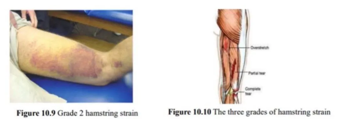
Mechanisms
| High-speed running | Dancing / kicking | |
|---|---|---|
| Movement | Generally believed to occur during the Terminal swing phaseThe last part of the swing phase, just before the foot makes contact with the ground.General meaning, not from the slides. In the second half of swing the hamstrings are actively lengthening and absorbing energy from the decelerating limb in preparation for foot contact | Slow or fast movements with simultaneous hip flexion and knee extension, placing the hamstrings in a position of extreme stretch |
| Muscle most affected | Biceps femorisThe hamstring muscle on the outer side of the back of the thigh.General meaning, not from the slides takes the greatest musculotendon stretch, which may contribute to its tendency to be more often injured than the other 2 hamstring muscles | SemimembranosusA hamstring muscle on the inner side of the back of the thigh.General meaning, not from the slides and its ProximalCloser to the trunk or to the point where a limb or structure attaches.General meaning, not from the slides free tendon (as opposed to the intramuscular tendon) |
- Extrinsic factors include training errors, such as increasing volume or intensity too quickly, particularly the sudden introduction of sprinting, lunging, hurdles or hills.
Examination
- Sudden onset of posterior thigh pain resulting from a specific activity, may be combined with an audible pop. Pain is more common at the proximal tendon.
- Individuals may also report pain at the Ischial tuberosityThe bony point at the bottom of the pelvis that you sit on. The hamstrings attach here.General meaning, not from the slides when sitting, most commonly when the proximal tendon is involved.
- Severe injuries (complete or partial ruptures of the hamstring muscles) typically result from extreme and forceful hip flexion with the knee fully extended (e.g. water skiing), and often require operative intervention with extensive postsurgical rehabilitation.
Hamstring syndromePain from sciatic nerve entrapment near the ischial tuberosity, where fibrous bands from the biceps insertion can form a tissue envelope around the nerve. vs complete or partial rupture. Both may produce similar pain at the ischial tuberosity, but the clinical presentation differs:
| Hamstring syndrome | Complete / partial rupture | |
|---|---|---|
| Sitting | Increased pain | Not provoked |
| SLRDIStraight leg raise, distal initiation: the straight leg raise test with ankle and foot dorsiflexion and the head and neck in neutral, so the test starts at the far end. / SDISlump distal initiation: the slump test started from the far end (distally), used to provoke nerve symptoms.General meaning, not from the slides | Could be provocative | Not provoked |
| Resisted knee flexion | Painful with hip flexed 90° and knee extended; pain free with hip extended (prone) | Painful with the hip both flexed and extended |
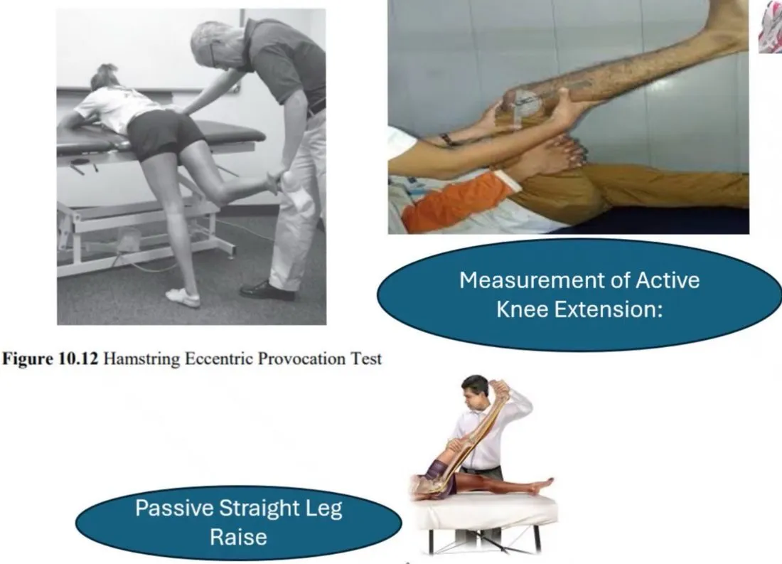
Special tests (slide 25)
The slide shows five tests as photos with labels: the hamstring EccentricA muscle contraction in which the muscle lengthens while producing force.General meaning, not from the slides provocation test (Figure 10.12), the resisted IsometricA muscle contraction in which the muscle stays the same length and the joint does not move.General meaning, not from the slides movement test, measurement of active knee extension, the passive Straight leg raiseLifting the straightened leg, either as a test (passive or active) or as a muscle test of the hip flexors.General meaning, not from the slides, and the Slump testA seated test that stretches the nerves along the back and legs, used to see whether neural tissue reproduces symptoms.General meaning, not from the slides.
Avulsions
- Proximal hamstring injuries should be evaluated with imaging techniques to rule out an Avulsion fractureFailure of the bone at the tendinous insertion rather than the tendon itself, so a piece of bone is pulled off..
- Proximal hamstring injuries typically include dramatic EcchymosisBruising: blood that has leaked under the skin.General meaning, not from the slides and swelling, with associated MorbidityIllness, disability or lasting problems caused by an injury.General meaning, not from the slides and a less positive PrognosisThe expected outcome of an injury or illness.General meaning, not from the slides.
- If a hamstring avulsion is complete, a PalpationExamining by feeling with the hands.General meaning, not from the slides gap is present at the hamstring attachment.
- As with all clinical findings, palpation findings become less valid as swelling and ecchymosis become present.
- Tendinous avulsions are most accurately diagnosed with MRIMagnetic resonance imaging: scans that show soft tissue and bone in detail without X-rays.General meaning, not from the slides.
- DistalFarther from the trunk or from the point where a limb or structure attaches.General meaning, not from the slides tears of hamstring tendons cause symptoms similar to those of typical hamstring strains at the Musculotendinous junctionThe point where muscle fibers blend into their tendon.General meaning, not from the slides of the biceps femoris.
- Reported symptoms: weakness and pain during knee flexion, a sense of instability of the knee joint, tenderness at the site of injury, and localized swelling and ecchymosis.
Rehabilitation of grade I and II hamstring strain
Three phases, each with goals, protection, ice, exercises, and criteria to move on.
| Phase 1 | Phase 2 | Phase 3 | |
|---|---|---|---|
| Goals | (1) Protect scar development · (2) Minimize AtrophyShrinking and wasting of a muscle.General meaning, not from the slides | (1) Regain pain-free hamstring strength, beginning in mid-range and progressing to a longer hamstring length · (2) Develop Neuromuscular controlHow well the nervous system coordinates muscles to produce steady, accurate movement.General meaning, not from the slides of trunk and pelvis with progressive increase in movement speed | (1) Symptom-free pain and tightness during all activities · (2) Normal ConcentricA muscle contraction in which the muscle shortens while producing force.General meaning, not from the slides and eccentric hamstring strength through full range of motion and speeds · (3) Improve neuromuscular control of trunk and pelvis · (4) Integrate postural control into sport-specific movements |
| Protection | Avoid excessive active or passive lengthening of the hamstrings. Static stretchingA stretch held in one position without movement.General meaning, not from the slides is not recommended (to avoid fiber disruption at this stage). Multi-angle isometrics should be initiated to align the regenerating muscle fibers and limit connective tissue FibrosisScar-like connective tissue forming in place of normal tissue.General meaning, not from the slides | Avoid end-range lengthening of hamstrings while hamstring weakness is present | Avoid full intensity if pain / tightness / stiffness is present |
| Ice | 2–3 times/d | Postexercise, 10–15 min | Postexercise, 10–15 min, as needed |
| Therapeutic exercise | Frequency not given on the slides | Performed 5–7 d/wk | Performed 4–5 d/wk |
Phase 1 is about protecting the healing muscle (no static stretching, isometrics to align the new fibers). Phase 2 rebuilds strength from mid-range toward longer lengths. Phase 3 returns full-range strength and sport-specific control.
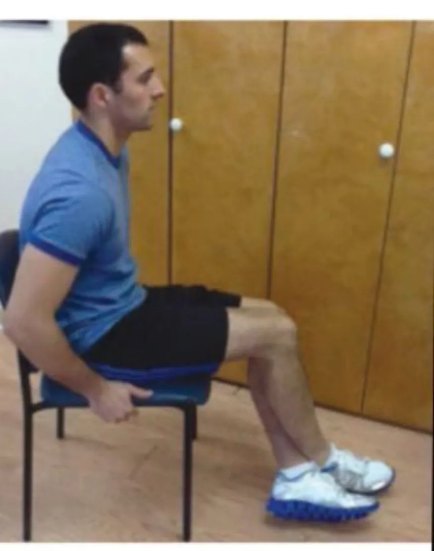
Phase 1 exercises
| # | Exercise | Dose |
|---|---|---|
| 1 | Stationary bike | × 10 min |
| 2 | Side-stepMoving sideways in small, quick steps.General meaning, not from the slides | × 10 m, 3 × 1 min, low to moderate intensity, pain-free speed and stride |
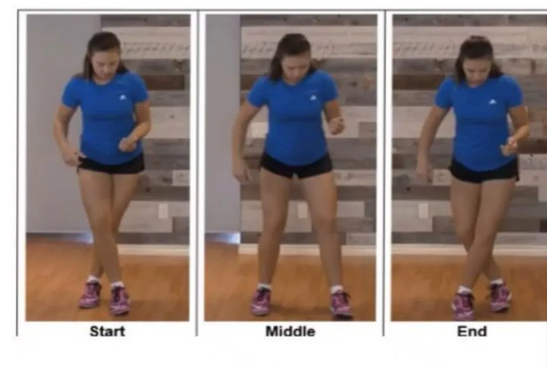
| 3 | GrapevineA sideways travelling step in which the trailing foot crosses in front of, then behind, the lead foot.General meaning, not from the slides | × 10 m, 3 × 1 min, low to moderate intensity, pain-free speed and stride |
| 4 | Fast feet steppingVery quick small steps on the spot.General meaning, not from the slides in place | 2 × 1 min |
| 5 | Prone body BridgeLifting the hips up from lying on the back, or holding a body-bridge position, to work the buttocks, hamstrings and trunk.General meaning, not from the slides | 5 × 10 s |
| 6 | Side body bridge | 5 × 10 s |
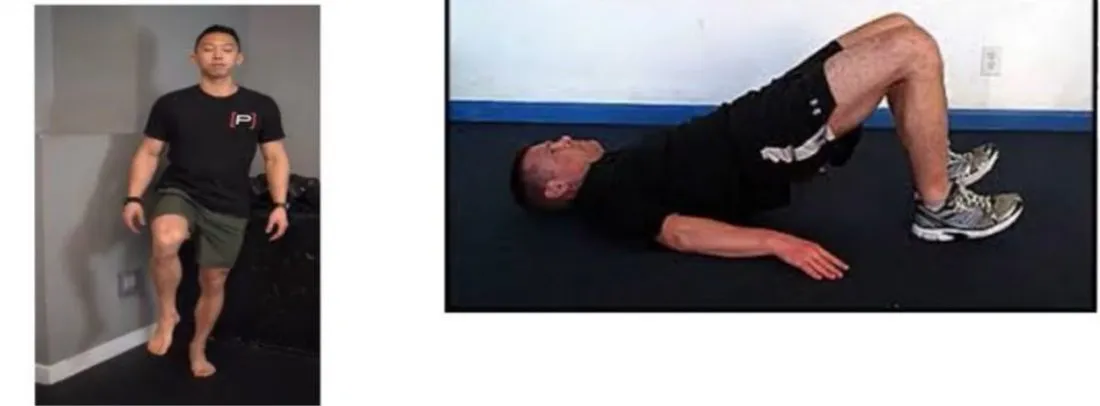
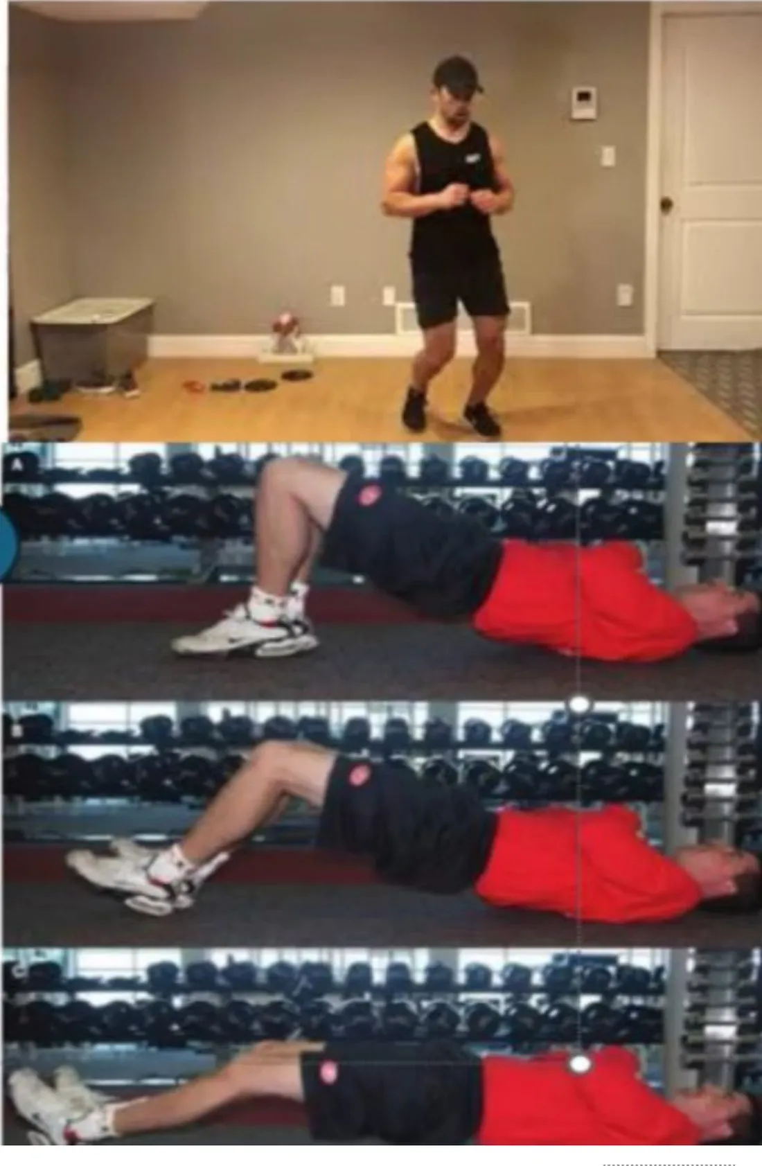
| 7 | Supine bent knee bridge | 10 × 5 s |
| 8 | Single-limb balance, progressing from eyes open to closed | 4 × 20 s |
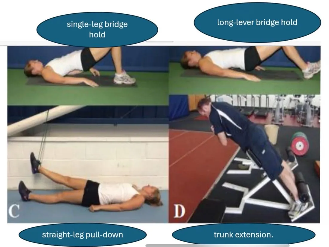
| 9 | Single-leg bridge hold, long lever bridge hold, straight-leg pull-down, trunk extension | no dose given on the slides |
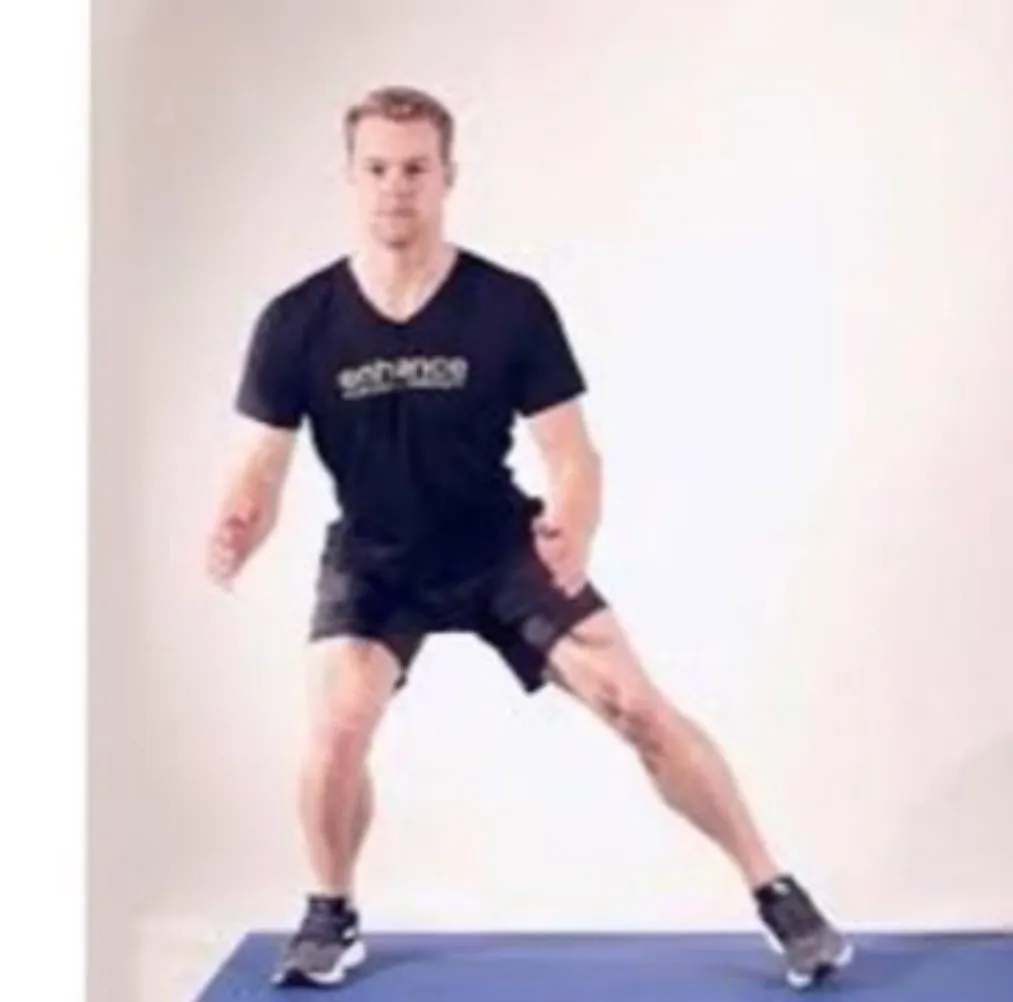


Criteria to progress from Phase 1 to Phase 2
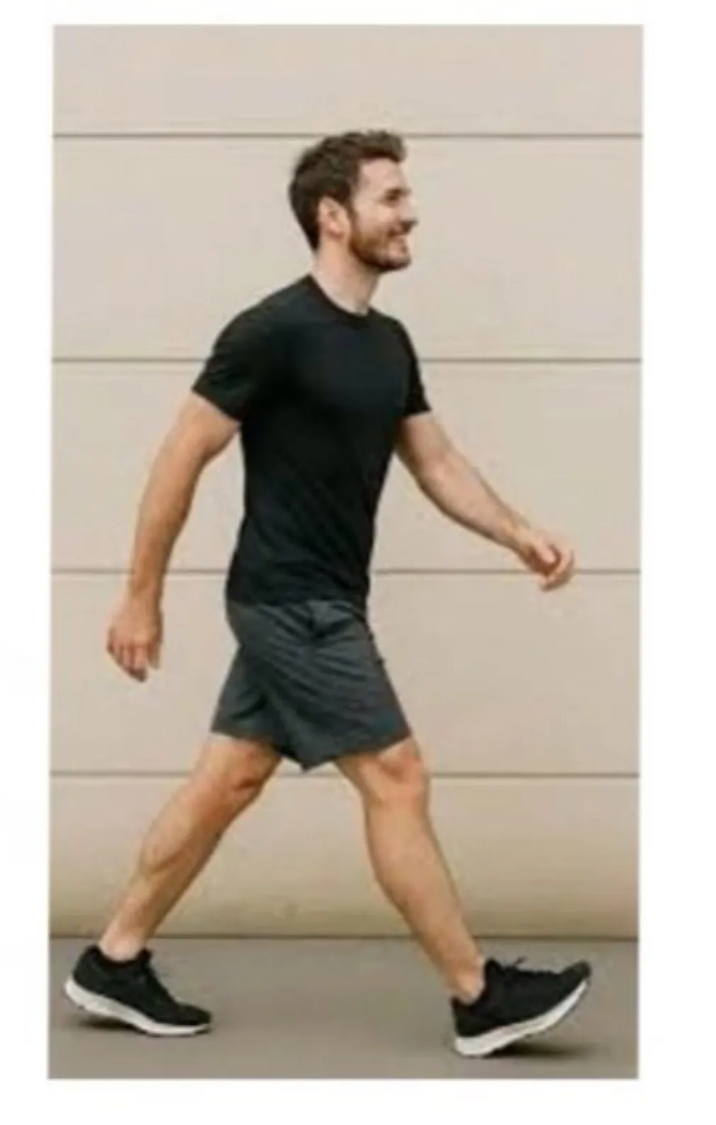
- Normal walking stride without pain.
- Very low-speed jog without pain.
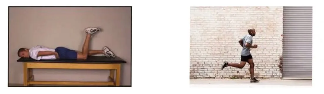
- Pain-free isometric contraction against submaximal (50%–70%) resistance during the prone knee flexion (90°) manual strength test.
Phase 2 exercises
| # | Exercise | Dose |
|---|---|---|
| 1 | Stationary bike | × 10 min |
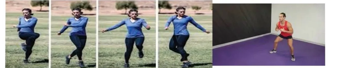
| 2 | Side-shuffle | × 10 m, 3 × 1 min, moderate to high intensity, pain-free speed and stride |
| 3 | Grapevine jog | × 10 m, 3 × 1 min, moderate to high intensity, pain-free speed and stride |
| 4 | Boxer shuffleA light, quick shuffling step from foot to foot, as a boxer moves.General meaning, not from the slides | × 10 m, 2 × 1 min, low to moderate intensity, pain-free speed and stride |
| 5 | Rotating body bridge | 5-s hold each side, 2 × 10 reps |
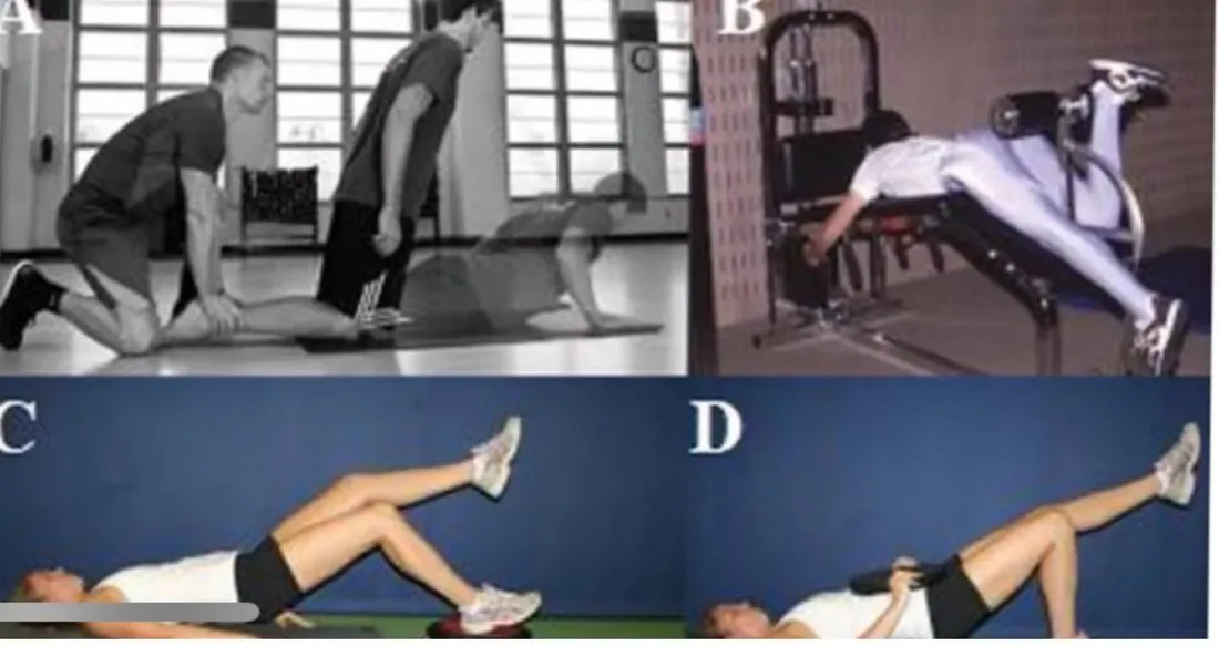
| 6 | Supine bent knee bridge with walk-outs | 3 × 10 reps |
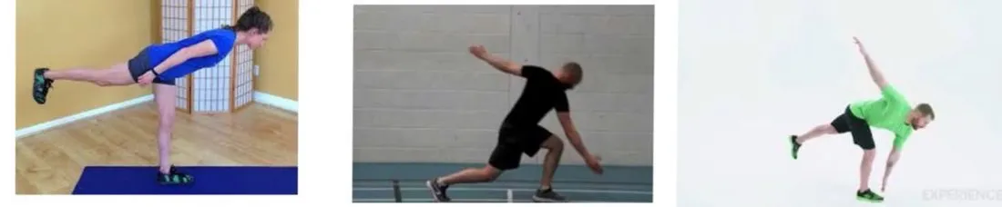
| 7 | Single-limb balance Windmill touchesBalancing on one leg while reaching down toward the floor with the opposite arm in a sweeping arc.General meaning, not from the slides without weight | 4 × 8 reps per arm each limb |
| 8 | LungeA step forward (or in another direction) with a deep bend of the front knee and hip.General meaning, not from the slides walk with trunk rotation, opposite hand-toe touch and T-lift | 2 × 10 steps per limb |
| 9 | Single-limb balance with forward trunk lean and opposite hip extension | 5 × 10 s per limb |
| 10 | Nordic hamstringA hamstring exercise in which the knees are held on the floor and the body is slowly lowered forward under control.General meaning, not from the slides, prone leg curl, supine leg curl, bridging progression | no dose given on the slides |
Criteria to progress from Phase 2 to Phase 3
- Full strength (5/5) without pain during the prone knee flexion (90°) manual strength test.
- Pain-free forward and backward jog, moderate intensity.
Phase 3 exercises
| # | Exercise | Dose |
|---|---|---|
| 1 | Stationary bike | × 10 min |
| 2 | Side-shuffle | × 30 m, 3 × 1 min, moderate to high intensity, pain-free speed and stride |
| 3 | Grapevine jog | × 30 m, 3 × 1 min, moderate to high intensity, pain-free speed and stride |
| 4 | Boxer shuffle | × 10 m, 2 × 1 min, moderate to high intensity, pain-free speed and stride |
| 6 | Forward-backward accelerations | 3 × 1 min; start at 5 m, progress to 10 m, then 20 m |
| 7 | Rotating body bridge with dumbbells | 5-s hold each side, 2 × 10 |
| 10 | Lunge walk with trunk rotation, opposite hand dumbbell toe touch and T-lift | 2 × 10 steps per limb |
| 11 | Sport-specific drills that incorporate postural control and progressive speed | no dose given on the slides |
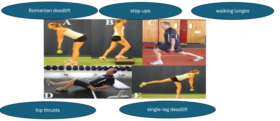
| 12 | Romanian deadliftA lifting exercise bending forward at the hips with the knees soft, to load the hamstrings and buttocks.General meaning, not from the slides, Step-upStepping up onto a raised surface, then back down.General meaning, not from the slides, walking lunges, Hip thrustDriving the hips up against resistance, with the upper back supported, to work the buttocks.General meaning, not from the slides, Single-leg deadliftA deadlift performed standing on one leg.General meaning, not from the slides | no dose given on the slides |
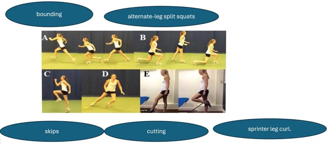
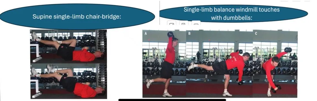
Pictured on slides 36 and 37 without an item number: BoundingExaggerated running strides that push off one leg and land on the other for distance.General meaning, not from the slides, Split squatA squat with the feet in a staggered stance, one foot forward and one back.General meaning, not from the slides, skips, CuttingMaking a sudden change of direction while running.General meaning, not from the slides, sprinter leg curl (slide 36), then supine single-limb chair-bridge and single-limb balance windmill touches with dumbbells (slide 37).
The slide numbering jumps 4 → 6 and 7 → 10. The unnumbered pictured exercises sit in those gaps in the slide order, but the slides do not number them.
Criteria for return to sport
- Full strength without pain:
- a. 4 consecutive repetitions of maximum effort manual strength test in each prone knee flexion position (90° and 15°).
- b. Less than 5% bilateral deficit in the eccentric hamstrings (30°/s) : concentric quadriceps (240°/s) ratio during IsokineticTesting or exercise on a machine that keeps the movement at a set speed.General meaning, not from the slides testing.
- c. Bilateral symmetry in the knee flexion angle of peak isokinetic concentric knee flexion torque at 60°/s.
- Full range of motion without pain.
- Replication of sport-specific movements near maximal speed without pain (e.g. incremental sprint test for running athletes).
Gluteal tendinopathy (Greater Trochanter Pain Syndrome)
Pathology
- Moderate to severe disabling pain over the Greater trochanterThe bony point on the outer side of the upper femur, where the gluteal muscles attach.General meaning, not from the slides (lateral hip pain) with marked tenderness over the greater trochanter.
- The combination of overuse and underlying weakness of the Gluteus mediusThe gluteal muscle on the outer side of the hip that abducts the hip and steadies the pelvis.General meaning, not from the slides and Gluteus minimusThe smallest gluteal muscle, lying under the gluteus medius.General meaning, not from the slides can cause StrainAn overstretch or tear of a muscle or its tendon. Graded I to III, where grade III is a complete tear.General meaning, not from the slides, tearing or degeneration of these muscles or their tendons, inducing TendinopathyPain and degeneration of a tendon, usually from overload.General meaning, not from the slides in the athlete.
- Tears of the gluteus medius and minimus may be a common cause of lateral trochanteric pain.
- PathomechanicsHow abnormal movement and loading lead to, or explain, an injury.General meaning, not from the slides: Gluteal tendinopathyTendon problem of the gluteus medius and minimus at the greater trochanter, causing lateral hip pain. The slide title also calls it greater trochanter pain syndrome. is often associated with Trochanteric bursitisInflammation of the bursa over the greater trochanter, the bony point on the outer side of the hip.General meaning, not from the slides, “the rotator cuff of the hip”, with regard to the broad origins and flat tendinous InsertionThe place where a tendon attaches to a bone.General meaning, not from the slides of the gluteals.
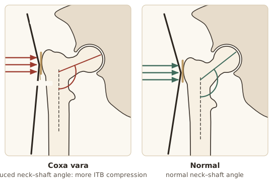
- Increased compressive force associated with a reduced Femoral neckThe narrower part of the femur between its head and shaft.General meaning, not from the slides–shaft angle is suggested to influence ITBIliotibial band: the thick band of tissue running down the outside of the thigh from the hip to the knee.General meaning, not from the slides compression against the gluteal tendons.
Clinical presentation
- Dull lateral hip pain, frequently aggravated by Weight-bearingPutting body weight through the leg or foot.General meaning, not from the slides activities such as walking, climbing stairs and running, and by resisted abduction.
- Pain can also occur when lying on the non-painful side if the painful hip falls into adduction (the hallmark sign for GT).
- Pain with prolonged sitting.
- Tenderness along the posterior aspect of the greater trochanter.
Examination
- There is no gold standard special test to rule in or rule out gluteal tendinopathy. The examination should include a thorough history, inspection, PalpationExamining by feeling with the hands.General meaning, not from the slides, range of motion, stability and assessment of strength in all planes.
- Regions and joints ProximalCloser to the trunk or to the point where a limb or structure attaches.General meaning, not from the slides (SIJSacroiliac joint: the joint between the sacrum and the hip bone (ilium) on each side of the pelvis.General meaning, not from the slides, LumbarRelating to the lower back, the part of the spine between the chest and the pelvis.General meaning, not from the slides, core/abdomen) and DistalFarther from the trunk or from the point where a limb or structure attaches.General meaning, not from the slides to the hip (knee, ankle, foot) should always be examined.
- Gait should be observed, noting Leg length discrepancyOne leg being shorter than the other.General meaning, not from the slides, compensations due to weakness, and heel strike and avoidance patterns.
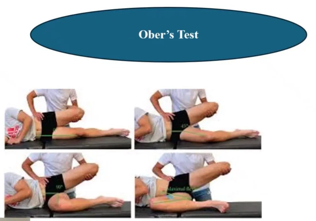
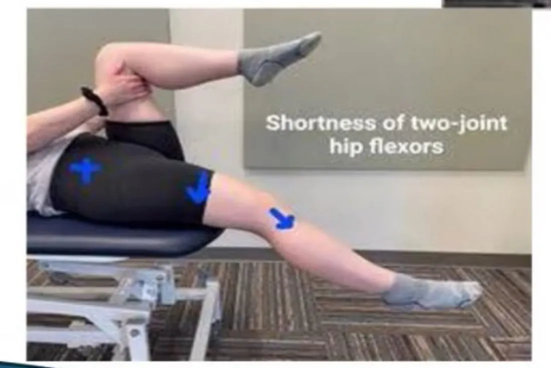
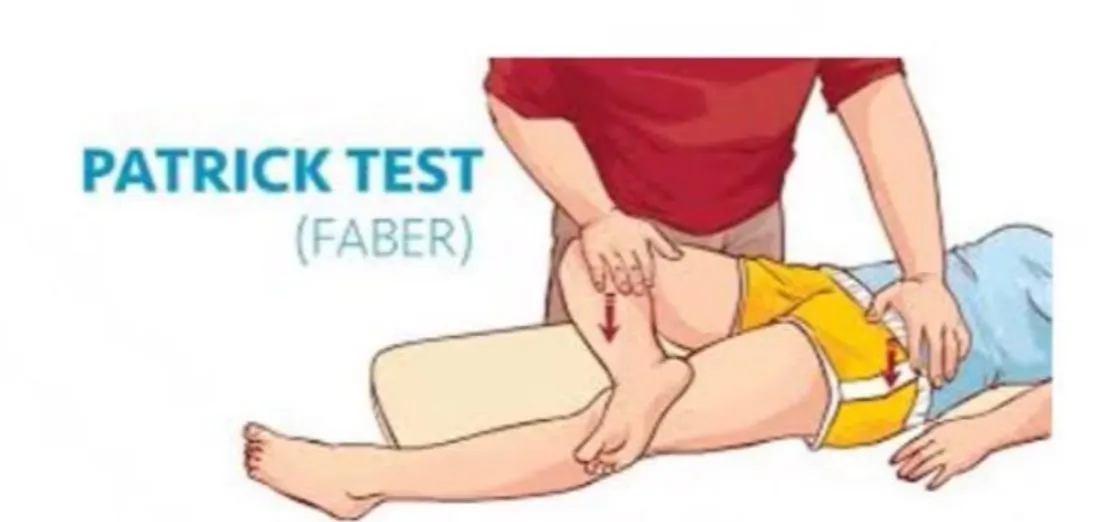
- Special tests to perform: Trendelenburg, Ober's testA side-lying test, often used to check tightness of the iliotibial band.General meaning, not from the slides, Thomas testA hip test pictured on the slide with the label shortness of two-joint hip flexors., FABERFlexion, abduction and external rotation of the hip, also called the Patrick test.General meaning, not from the slides and Ely's testA test, done lying face down with the knee bent, for tightness of the rectus femoris.General meaning, not from the slides tests.
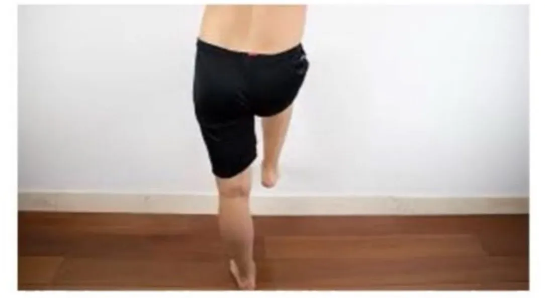
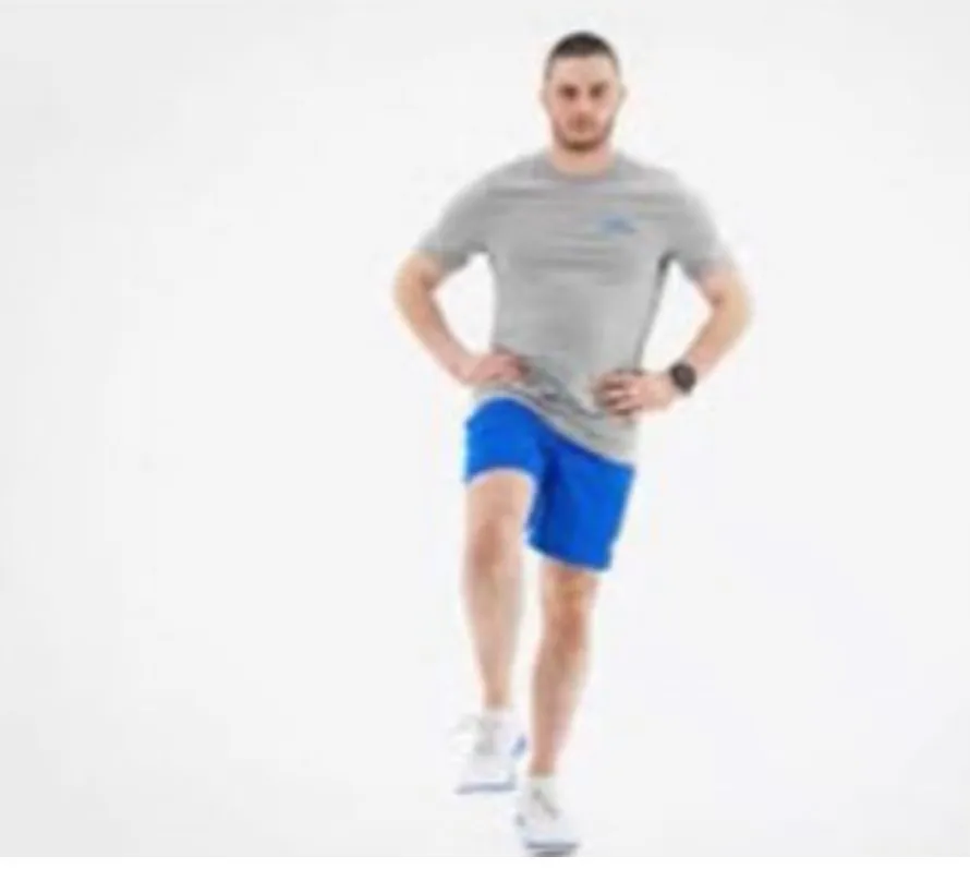
- The Trendelenburg testA standing single-leg test. The slide says it assesses the functional strength of the gluteus medius. assesses the functional strength of the gluteus medius.
Slide 43 shows four of these tests: Ober’s test, Trendelenburg test, Thomas test (the picture is labelled “shortness of two-joint Hip flexorsThe muscles at the front of the hip that lift the thigh toward the trunk.General meaning, not from the slides”) and the Patrick test (FABER). Ely’s test is listed on slide 42 but has no picture.
Four tests with their values (slide 44)
| # | Test | What the slide says |
|---|---|---|
| 1 | Palpation | Most sensitive (0.83) but least specific (0.43) |
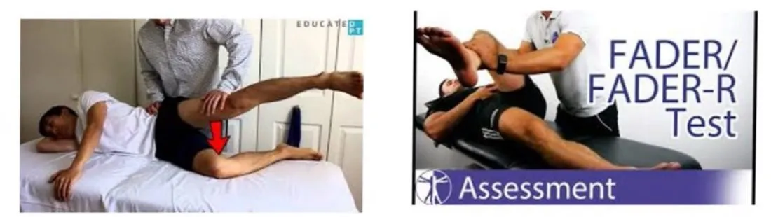
| 2 | FADERFlexion, adduction and external rotation: the therapist passively takes the limb into 90° hip flexion, adduction and external rotation to end of range. (Flexion / Adduction / External Rotation) | The therapist passively takes the limb into 90° hip flexion, adduction and external rotation to end of range. It aims to position the ITB over the greater trochanter, and puts the gluteus medius and minimus tendons under tension with the ITB fascia against the greater trochanter |
| 3 | Resisted hip abduction | 71% SensitivityHow often a test is positive in people who really have the condition.General meaning, not from the slides and 84% SpecificityHow often a test is negative in people who do not have the condition.General meaning, not from the slides. Assessing active abduction in a position of hip adduction may be more useful |
| 4 | 30-second single leg stance test | The person must stand, unassisted, on one leg with eyes open and one finger on a wall. Specificity 100%, sensitivity 38% and Positive predictive valueThe chance that a positive test result is correct.General meaning, not from the slides 100% |
The slide’s sentence on the single leg stance test stops at “indicating that if the test was positive” and the rest is missing.
Rehabilitation program
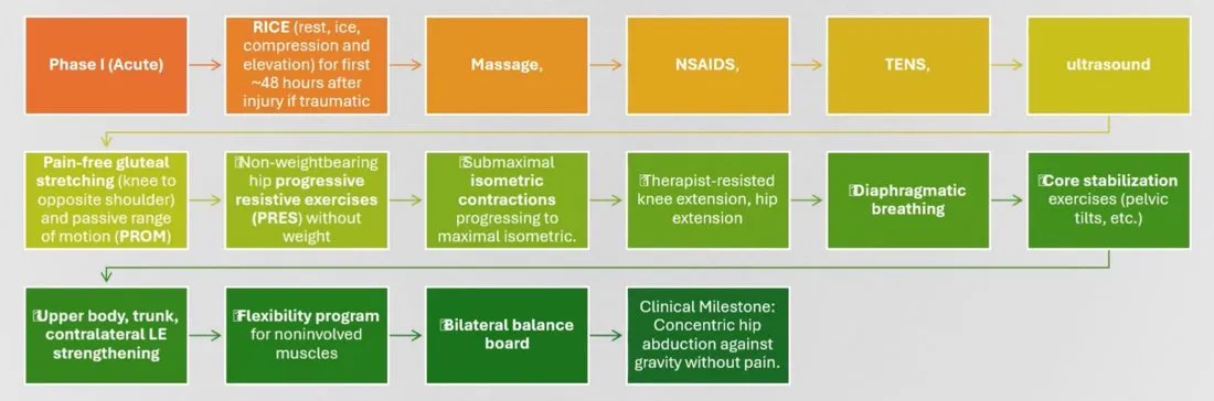
Phase I (Acute)
Flowchart on slide 45, in order:
- RICERest, ice, compression and elevation. (rest, ice, compression and elevation) for the first ~48 hours after injury if traumatic.
- Massage, NSAIDsNonsteroidal anti-inflammatory drugs, such as ibuprofen.General meaning, not from the slides, TENSTranscutaneous electrical nerve stimulation: mild electrical current through the skin to ease pain.General meaning, not from the slides, UltrasoundSound waves used either as a treatment (heating tissue) or to produce images.General meaning, not from the slides.
- Pain-free gluteal stretching (knee to opposite shoulder) and PROMPassive range of motion: how far a joint moves when the therapist moves it, with no effort from the patient.General meaning, not from the slides (PROM).
- Non-weightbearing hip PRESProgressive resistive exercises: strengthening exercises where the resistance is raised step by step.General meaning, not from the slides (PRES) without weight.
- Submaximal IsometricA muscle contraction in which the muscle stays the same length and the joint does not move.General meaning, not from the slides contractions progressing to maximal isometric.
- Therapist-resisted knee extension, hip extension.
- Diaphragmatic breathingDeep breathing that uses the diaphragm so the belly rises as you breathe in.General meaning, not from the slides.
- Core stabilizationExercises that train the trunk muscles to hold the spine and pelvis steady.General meaning, not from the slides exercises (Pelvic tiltRocking the pelvis forward or backward to change the curve of the lower back.General meaning, not from the slides, etc.).
- Upper body, trunk, ContralateralOn the opposite side of the body.General meaning, not from the slides LELower extremity: the leg.General meaning, not from the slides strengthening.
- Flexibility program for noninvolved muscles.
- Bilateral Balance boardAn unstable board used to train balance and joint control.General meaning, not from the slides.
- Clinical milestone: ConcentricA muscle contraction in which the muscle shortens while producing force.General meaning, not from the slides hip abduction against gravity without pain.
Phase II (Subacute)
- Bicycling / elliptical / stair master / swimming.
- Non-weightbearing hip PRES with weight.
- BridgeLifting the hips up from lying on the back, or holding a body-bridge position, to work the buttocks, hamstrings and trunk.General meaning, not from the slides (bilateral progressing to unilateral).
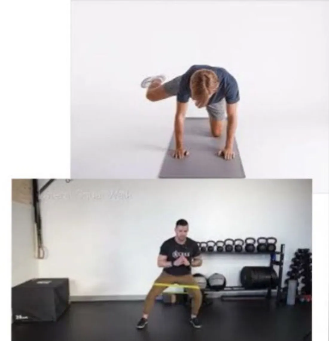
- QuadrupedOn hands and knees.General meaning, not from the slides hip extension, Fire hydrantOn hands and knees, lifting the bent knee out to the side.General meaning, not from the slides.
- PlankA hold supported on the forearms or hands and the feet, with the body kept in a straight line.General meaning, not from the slides, side planks.
- Weight-bearing hip PRES with TBTheraband: an elastic resistance band used for strengthening.General meaning, not from the slides (TB) or cable column (concentric to EccentricA muscle contraction in which the muscle lengthens while producing force.General meaning, not from the slides).
- TB SquatBending the hips and knees to lower the body, then standing back up.General meaning, not from the slides, lateral walks.
- Single limb stance, step down, LungeA step forward (or in another direction) with a deep bend of the front knee and hip.General meaning, not from the slides with reciprocal arm movements.
- Eccentric short lever ERExternal rotation: turning the limb outward.General meaning, not from the slides fall outs.
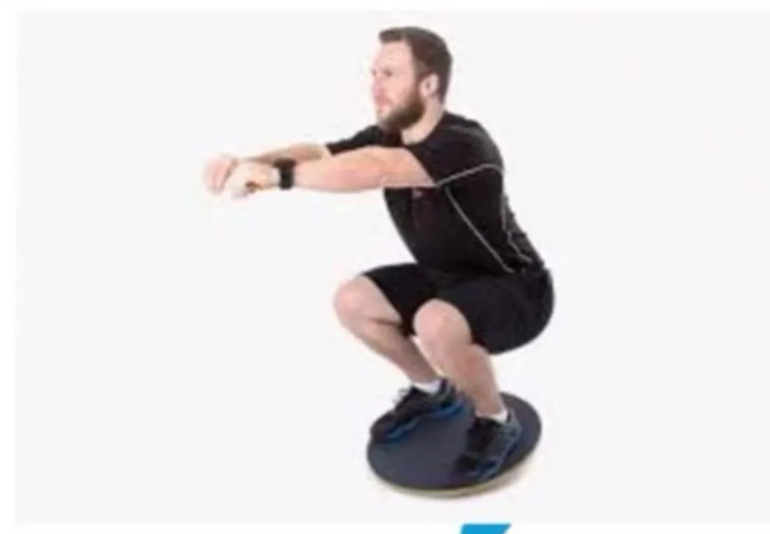
- Balance board squats with throwbacks.
- General flexibility program.
- Clinical milestone: ability to perform pain-free short lever eccentric training.
Phase III (Sports specific training)
- Phase II exercises with increased load, intensity, speed and volume.
- Plank progression: prone plank + hip extension, side plank + abduction with ER.
- Standing resisted stride lengths on cable column.
- Slide board.
- Lunges (in all planes) with progressing balance challenges (AerofoamA soft foam pad that creates an unstable surface for balance training.General meaning, not from the slides → BOSUA half-dome balance trainer, used flat side up or dome up.General meaning, not from the slides).
- Progressive balance exercises.
- Standing hip ER → standing hip ER + TB unilateral squat.
- Eccentric training: standing TB eccentric, treadmill resistance, Step-upStepping up onto a raised surface, then back down.General meaning, not from the slides plus eccentrics.
- PlyometricFast, spring-like exercises such as jumping that quickly stretch and then shorten a muscle.General meaning, not from the slides sport-specific training.
- Correct or modify sport-specific movements.
- Clinical milestone: pain-free sport-specific activities.
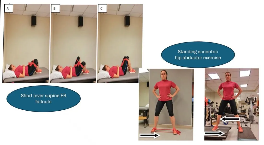
Each phase ends in a clinical milestone: Phase I = concentric hip abduction against gravity without pain; Phase II = pain-free short lever eccentric training; Phase III = pain-free sport-specific activities.
Groin pain
- The most common cause of groin pain is increased tendon load during directional-change types of sports, associated with stress shielding in the InsertionThe place where a tendon attaches to a bone.General meaning, not from the slides of the tendon where collagen gradually transitions to mineralized cartilage and bone. However, there is controversy as to what structures are most responsible under this loaded condition.
- Slide wording of the second bullet: “TendinopathyPain and degeneration of a tendon, usually from overload.General meaning, not from the slides is most responsible for the pain, Osteitis pubisInflammation of the pubic symphysis with bone changes around it, giving groin and pubic pain that is frequently severe., a Athletic pubalgiaAlso called sport's hernia or hockey hernia: weakening or tearing of the abdominal wall structures, creating an inside-out hernia in the dorsal wall of the inguinal canal.”.
The second bullet is incomplete on the slide. It reads like a list of three candidate causes of groin pain: tendinopathy, osteitis pubis, sportsman’s hernia (both are covered later in this lecture).
Adductor tendinopathies
| Structure | Most painful with |
|---|---|
| Adductor longusA thigh muscle on the inner side that pulls the leg toward the midline. The slide groups it with the adductor brevis.General meaning, not from the slides and brevis | Resisted hip adduction with the hip positioned in neutral |
| GracilisA long, thin inner-thigh muscle that adducts the hip and also flexes the knee.General meaning, not from the slides | Resisted hip adduction in the same position (neutral), along with painful resisted knee flexion |
| PectineusA small muscle at the front of the upper inner thigh that flexes and adducts the hip.General meaning, not from the slides | Resisted hip flexion and resisted hip adduction with the hip positioned in 90° of flexion |
| Rectus abdominisThe straight muscle at the front of the abdomen that flexes the trunk.General meaning, not from the slides tendon | Implicated as a source of chronic groin pain during resisted hip adduction, since it attaches at the superior surface of the Pubic tubercleA small bony bump on the upper edge of the pubic bone.General meaning, not from the slides. The patient performs resisted trunk flexion in the supine position |
- In rare cases, long-standing pain may suggest Myositis ossificansBone-like tissue forming inside a muscle, usually after an injury.General meaning, not from the slides of the adductors, when symptoms persist after trauma and resist conservative management (the slide adds “imaging”).
Treatment of adductor tendinopathies
The slide title reads only “TTT”.
- Tendinopathies can be treated with strengthening and/or Transverse friction massageFirm massage applied across the fibers of a tendon or muscle at the injured spot.General meaning, not from the slides at the insertion into the pubic tubercle, stretching, and gradual strengthening that leads to a return to activity.
- The efficacy of transverse friction massage has been questioned, but it is one treatment option.
- Friction massage has been found to stimulate FibroblastA cell that makes collagen and builds new tissue during healing.General meaning, not from the slides proliferation and promote tissue healing through increased fibroblast recruitment.
- It can produce a temporary AnalgesicsPain-relieving medicines, or the state of pain relief.General meaning, not from the slides through application of a noxious stimulus that requires approximately two minutes to reach a pain-free threshold, lending hours or days of relief.
- If the affliction involves muscle tissue, transverse friction massage has been shown to increase blood flow to the area where the friction is applied.
- When transverse friction fails, the tendinopathy is treated with injection at the bony insertion, which might be best guided with FluoroscopyLive X-ray imaging, used here to guide a needle to the right place.General meaning, not from the slides assistance.
- Fluoroscopically guided injection might be best suited for Iliopectineal bursitisInflammation of the iliopectineal bursa, a cushion at the front of the hip next to the iliopsoas tendon.General meaning, not from the slides, which can produce symptoms likened to IliopsoasThe combined psoas and iliacus muscles, the main hip flexors.General meaning, not from the slides tendinopathy. This condition presents with passive hip tests most painful: passive hip extension, flexion and external rotation in a position of full hip flexion produce the greatest symptoms (due to compression of the Lesser trochanterThe small bony point on the inner upper femur, where the iliopsoas attaches.General meaning, not from the slides against the BursaA small fluid-filled sac that cushions the space between a bone and a tendon or muscle.General meaning, not from the slides).
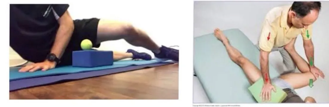
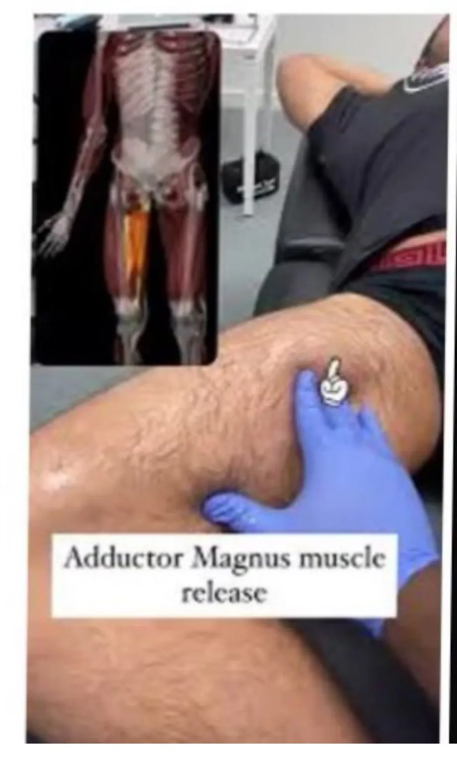
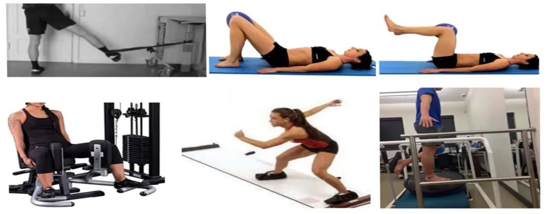
Strengthening exercises for Adductor tendinopathyPain and degeneration of the tendons of the inner thigh (adductor) muscles, usually felt in the groin.General meaning, not from the slides (slide 52, photos only).
Osteitis pubis
- Severe pubic pain can also be caused by a pubic bone stress injury and is often associated with athletic trauma (this first sentence is struck through with a pen line on the slide).
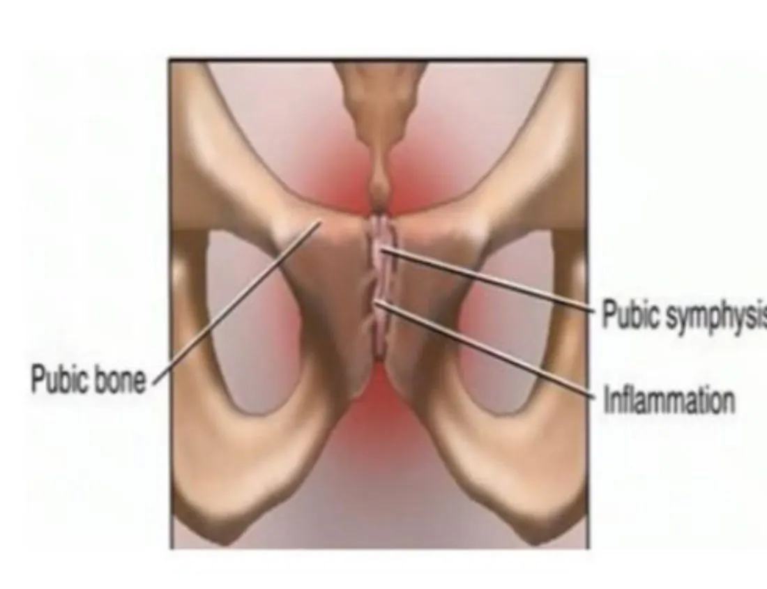
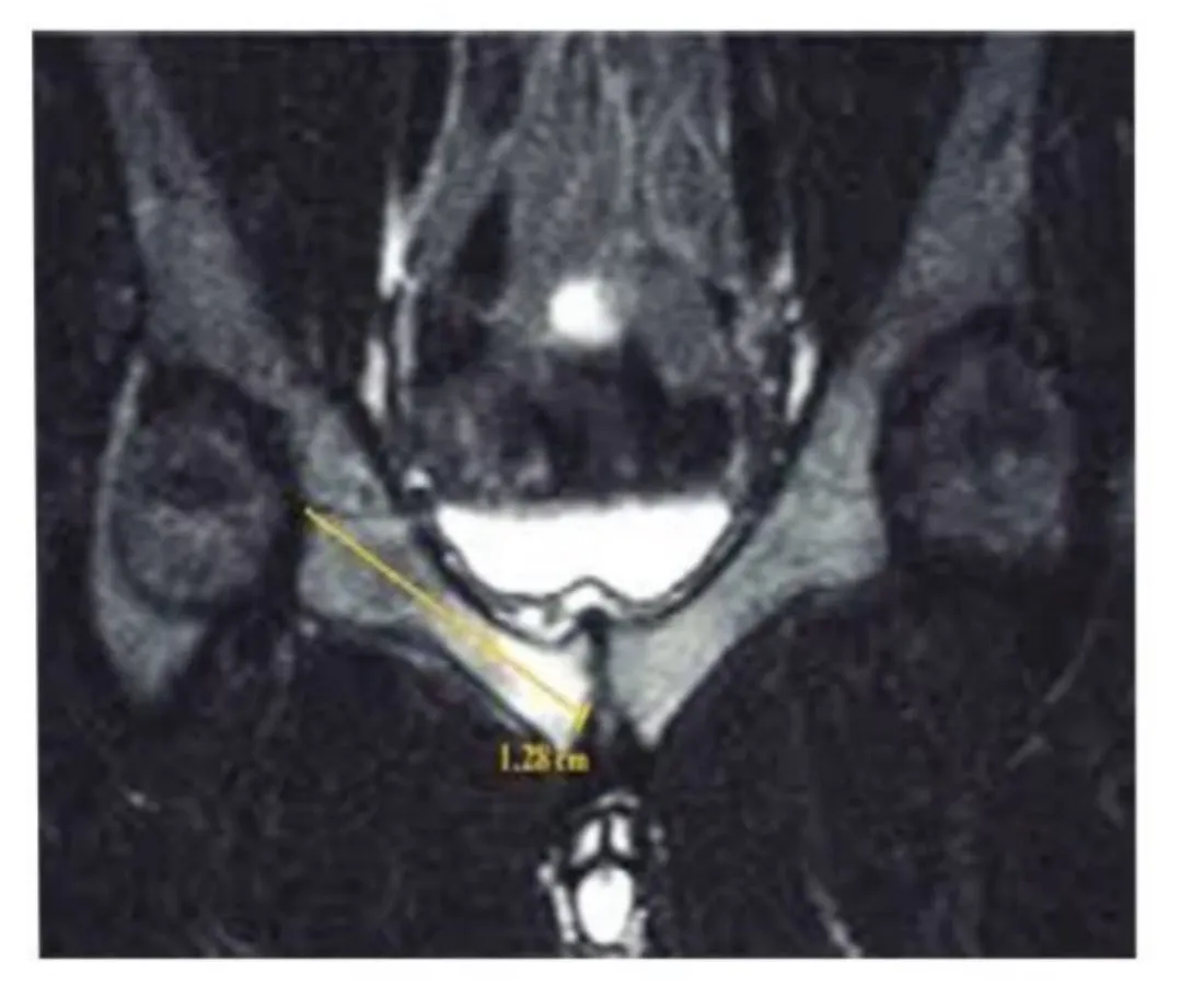
- Osteitis pubis produces symphyseal inflammation, periarticular bone ScleroticHardened or denser than normal, as bone can become.General meaning, not from the slides changes, and resultant groin and pubic pain that is frequently severe.
- Accompanying ParasymphysealNext to the pubic symphysis.General meaning, not from the slides Bone marrow edemaFluid-like swelling within the bone marrow, visible on MRI.General meaning, not from the slides can be seen on T2 MRIAn MRI image type in which fluid, such as swelling or edema, shows up bright.General meaning, not from the slides, which appears to strongly correlate with the clinical presentation.
- Conservative treatment typically fails, thus requiring more aggressive management strategies. Topol et al. reported promising results with ProlotherapyA treatment that involves injecting injuries with irritant solutions, thereby stimulating healing and strengthening of tissue., a treatment that involves injecting injuries with irritant solutions, thereby stimulating healing and strengthening of tissue.
- Finally, surgical stabilization of the symphysis has been used when other conservative forms of treatment are unsuccessful.
Athletic pubalgia (sport’s hernia)
Also labelled “hockey hernia” or “athletic pubalgia”.
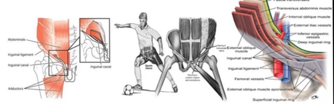
- Patients with abdominal musculoskeletal abnormalities experience a weakening or tearing of the Transversalis fasciaA thin sheet of connective tissue lining the inside of the abdominal wall.General meaning, not from the slides, Conjoined tendonA tendon formed where fibers of two abdominal muscles join, attaching at the pubic bone.General meaning, not from the slides, and/or Internal obliqueAn abdominal muscle on the side of the trunk that helps rotate and bend the trunk.General meaning, not from the slides fibers, creating an inside-out HerniaA bulge of tissue pushing through a weak spot in the wall that normally holds it in.General meaning, not from the slides within the dorsal wall of the Inguinal canalA passage through the lower abdominal wall in the groin region.General meaning, not from the slides.
- Causes: this traumatic injury has been associated with twisting, turning, or directional changes in speed causing the hip to move into abduction, adduction or extension. BallisticFast, explosive movements.General meaning, not from the slides movements, as frequently observed in soccer and ice hockey players, lead to shearing at the Symphysis pubisThe joint at the front of the pelvis where the two pubic bones meet.General meaning, not from the slides and resultant stress on the above structures.
Clinical findings (slide 55)
- Can cause debilitating pain in the lower abdominal, inguinal and groin regions, either unilateral or bilateral, exacerbated with exertion or ValsalvaBearing down or straining with the airway closed, as when lifting or coughing hard.General meaning, not from the slides.
- Resisted double hip adduction will be painful and unchanged with the Pelvic ring stabilization beltA belt placed around the pelvis to hold the pelvic ring steady while testing.General meaning, not from the slides.
- Resistive hip tests may be weak, and resisted oblique abdominal tests will be painful and possibly weak when the hip is positioned in extension.
- PalpationExamining by feeling with the hands.General meaning, not from the slides of the adductors is often unprovocative.
- Plain film imaging might reveal an associated osteitis pubis, but diagnostic UltrasoundSound waves used either as a treatment (heating tissue) or to produce images.General meaning, not from the slides tends to provide the best support to the diagnosis.
- A strong clinical suspicion could merit surgical exploratory evaluation in the absence of true findings on imaging.
- Patients who fail conservative management may be candidates for surgical intervention.
Five signs indicative of athletic pubalgia
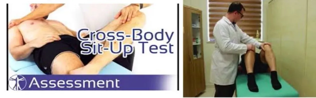
- A subjective complaint of deep groin / lower abdominal pain.
- Pain that is exacerbated with sport-specific activities such as sprinting, kicking, CuttingMaking a sudden change of direction while running.General meaning, not from the slides and/or sit-ups, and is relieved with rest.
- Palpable tenderness over the Pubic ramusThe arm of the pubic bone that extends out from the pubic symphysis.General meaning, not from the slides at the insertion of the rectus abdominis and/or conjoined tendon.
- Pain with resisted hip adduction at 45° of hip flexion.
- Pain with resisted abdominal curl-up.
Surgical repair
- Surgical LaparoscopicKeyhole surgery done through small cuts, using a camera.General meaning, not from the slides of the defect has been successful. 97.1% return to sport after laparoscopic surgical repair of the defect.
- However, Open mesh repairOpen surgery in which a mesh patch is used to reinforce the weak area.General meaning, not from the slides appears superior to laparoscopic repair.
Labral tears
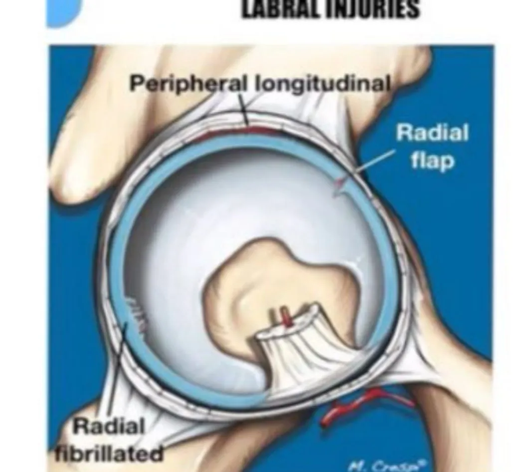
Causes and classification
- Labral tearA tear in the labrum of the hip. The slides classify tears as traumatic vertical or degenerative horizontal.General meaning, not from the slides can result from micro or macrotrauma, degeneration related to DysplasiaAbnormal development of a joint, such as a socket that is too shallow.General meaning, not from the slides or CFJCoxafemoral joint: the hip joint, where the head of the femur sits in the socket of the pelvis.General meaning, not from the slides instability, and developmental deficiencies.
- Labral lesions are more common between the ages of 18 and 40.
- Acetabular labral tears may occur as the result of acute trauma or of insidious onset.
- Traumatic mechanisms involve rapid twisting, pivoting or falling motions.
- A common mechanism in the athletic population includes forceful rotation with the hip in a hyperextended position.
- The Modified circumduction testA hip test labelled on the slides as a hip impingement test. It is used as a provocative maneuver for labral tears. can be used as a provocative maneuver.
- Labral tears should be Differential diagnosisSorting through the possible causes of a symptom to work out which one fits best.General meaning, not from the slides from hip Hip chondromalaciaSoftening and damage of joint cartilage.General meaning, not from the slides, Loose bodiesSmall fragments of bone or cartilage floating free inside a joint.General meaning, not from the slides and Internal snapping hipA snapping or clicking sensation in the hip during movement, of the internal type.General meaning, not from the slides.
- A FluoroscopyLive X-ray imaging, used here to guide a needle to the right place.General meaning, not from the slides guided IntraarticularInside the joint.General meaning, not from the slides injection can be used to differentiate symptoms that are intraarticular versus ExtraarticularOutside the joint, in the tissues around it.General meaning, not from the slides.
- Labral tears can be classified as either traumatic vertical or degenerative horizontal tears.
- Vertical traumatic tears can be classified as either full substance or partial substance with Avulsion fractureFailure of the bone at the tendinous insertion rather than the tendon itself, so a piece of bone is pulled off..
Pathology and clinical picture
- This injury is thought to be similar to a SLAP lesionSuperior labrum anterior to posterior lesion: a tear of the labrum at the top of the shoulder socket.General meaning, not from the slides of the GlenohumeralRelating to the shoulder joint, where the humerus meets the shoulder blade.General meaning, not from the slides LabrumThe ring of cartilage around the rim of the hip socket (acetabulum). It deepens the socket and helps seal the joint.General meaning, not from the slides.
- Tears most frequently occur in the anterior superior or posterior superior margins of the labrum, due to compromised mechanical properties, increased mechanical demand, poor vascular penetration, and impact loading of the Femoral headThe ball-shaped top of the thigh bone that sits in the hip socket.General meaning, not from the slides–neck junction against the labral rim.
- A torn labrum can be related to dysplasia, promotes increased joint instability, and serves as a precursor to CFJ ArthrosisWear and degeneration of a joint.General meaning, not from the slides.
- The patient often complains of Mechanical symptomsLocking, clicking, catching and giving way, as listed on the slides. such as locking, clicking, catching and giving way.
- Pain is usually reported within the anterior hip or in the anterior groin region, although some patients report pain in the posterior buttock.
Conservative management and surgery (slide 58)
- Athletes with labral pathology may respond to conservative management, and this should always be trialed prior to undergoing surgery.
- Repetitive hip flexion, adduction or abduction and rotation at the end of range should be avoided through activity modification.
- High velocity, low amplitudeA mobilization or manipulation technique using a quick, small-range movement.General meaning, not from the slides rotational mobilization/manipulation can be attempted, as long as Necrotic changesChanges caused by the death of tissue (necrosis).General meaning, not from the slides have been ruled out.
- If conservative measures fail to reduce the symptoms, then ArthroscopicDone through small cuts using a camera inside the joint (keyhole surgery).General meaning, not from the slides excision and partial LabrectomySurgical removal of part of the labrum.General meaning, not from the slides can be implemented.
Femoroacetabular impingement (FAI)
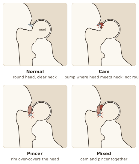
In Femoroacetabular impingementAn abnormal shape of the proximal femur and acetabulum that raises contact forces in hip flexion, potentially injuring the labrum and cartilage and causing pain., an abnormal morphology of the ProximalCloser to the trunk or to the point where a limb or structure attaches.General meaning, not from the slides femur and AcetabulumThe cup-shaped socket of the hip joint, in the pelvis.General meaning, not from the slides results in increased hip contact forces with hip motion in hip flexion, thus potentially leading to LabrumThe ring of cartilage around the rim of the hip socket (acetabulum). It deepens the socket and helps seal the joint.General meaning, not from the slides-cartilage injury and pain.
| Cam impingementImpingement linked to a non-spherical femoral head and neck relationship. The loss of roundness causes abnormal contact between the head and socket. | Pincer impingementImpingement where the acetabulum has too much coverage around the femoral head, so the labral cartilage is pinched against the femoral head-neck junction. | Mixed impingementCam and pincer impingement existing together. | |
|---|---|---|---|
| What it is | Associated with a non-spherical Femoral headThe ball-shaped top of the thigh bone that sits in the hip socket.General meaning, not from the slides and neck relationship. This loss of roundness contributes to abnormal contact between the head and socket | The acetabulum has too much coverage around the femoral head, commonly along the front-top rim of the socket, so the labral cartilage is “pinched” between the rim of the socket and the anterior femoral head–neck junction | Cam and pincer coexist (frequently) |
| Typically secondary to | — | “RetroversionA turning back of the acetabulum, one of the shapes behind pincer impingement.” (a turning back of the acetabulum), “ProfundaA socket that is too deep, one of the shapes behind pincer impingement.” (a socket that is too deep), or “ProtrusioA situation where the femoral head extends into the pelvis.” (the femoral head extends into the pelvis) | — |
Clinical findings
- Pain in the anterior hip / groin and/or lateral hip / trochanter region.
- Pain is described as aching or sharp.
- Pain is aggravated by sitting.
- Pain is reproduced with the hip flexion, adduction and internal rotation (FADIRFlexion, adduction and internal rotation: the hip position whose pain reproduction the slide lists as a finding in femoroacetabular impingement.) test.
- Hip flexion and abduction are limited.
- Mechanical symptomsLocking, clicking, catching and giving way, as listed on the slides. such as popping, locking or snapping.
- FAI can be identified using FAI testing.
- A patient with a Labral tearA tear in the labrum of the hip. The slides classify tears as traumatic vertical or degenerative horizontal.General meaning, not from the slides will complain of increased pain with sitting and climbing stairs, along with possible clicking, locking and/or giving way during Weight-bearingPutting body weight through the leg or foot.General meaning, not from the slides activities.
- The patient will present with pain and possible limitation during passive internal rotation with the hip flexed, but not with the hip extended.
- The pain will increase with the Modified circumduction testA hip test labelled on the slides as a hip impingement test. It is used as a provocative maneuver for labral tears..
- Magnetic resonance arthrographyAn MRI scan done after contrast dye is injected into the joint, to show the cartilage and labrum more clearly.General meaning, not from the slides can help confirm the diagnosis.
Conservative treatment of labral lesions
The slide title reads “TTT” again.
- Education and participation in unloaded activities for the hip.
- Instruct the patient to change seating surfaces so that seated hip flexion is reduced.
- Teach the patient to reduce extensive sitting and the use of stairs.
- Initially treat with supported ambulation using an Assistive deviceAn aid such as a cane, crutches or a walker.General meaning, not from the slides.
- Cycling can be continued, as long as the load is significantly reduced.
- High velocity, low amplitudeA mobilization or manipulation technique using a quick, small-range movement.General meaning, not from the slides rotational mobilization/manipulation can be attempted, as long as Necrotic changesChanges caused by the death of tissue (necrosis).General meaning, not from the slides have been ruled out.
- IntraarticularInside the joint.General meaning, not from the slides injection of CorticosteroidsStrong anti-inflammatory medicines, here given by injection.General meaning, not from the slides can reduce the inflammation and CytokinesChemical messengers released by cells that drive inflammation.General meaning, not from the slides proliferation associated with a chronic condition.
- Steroid injections and CT-guided aspirationUsing CT scan images to guide a needle that draws out fluid.General meaning, not from the slides may be effective if cystic changes within the labrum are present.
- If conservative measures fail, ArthroscopicDone through small cuts using a camera inside the joint (keyhole surgery).General meaning, not from the slides excision and partial LabrectomySurgical removal of part of the labrum.General meaning, not from the slides can be implemented.
- In extreme conditions, surgeons may elect to perform an Acetabular derotation osteotomySurgery that cuts the pelvic bone around the socket and turns it to change the socket's position.General meaning, not from the slides to reduce the stress of the Femoral neckThe narrower part of the femur between its head and shaft.General meaning, not from the slides on the labrum.
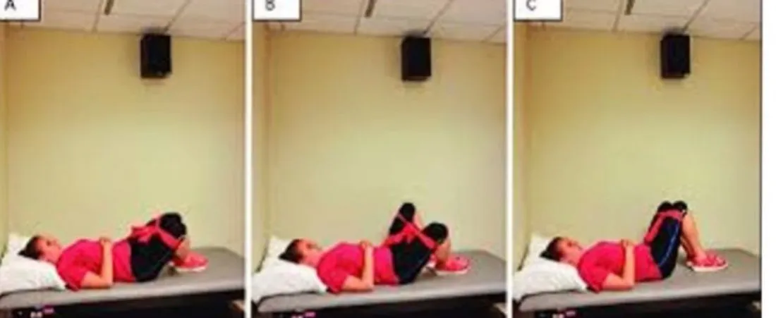
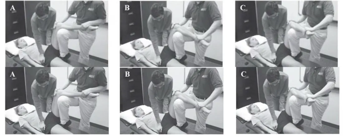
Labral manipulation (slide 63, photo sequences A–C shown twice).
Avulsion fractures and apophyseal injuries
- In the hip and pelvis, they result from failure of the bone at the tendinous InsertionThe place where a tendon attaches to a bone.General meaning, not from the slides rather than the tendon itself.
- More common in skeletally immature athletes with open ApophysisA growth area of bone where a tendon attaches. It is still open in skeletally immature athletes.General meaning, not from the slides.
- Usually the result of a sudden, forceful ConcentricA muscle contraction in which the muscle shortens while producing force.General meaning, not from the slides or EccentricA muscle contraction in which the muscle lengthens while producing force.General meaning, not from the slides contraction or rapid, excessive passive lengthening.
- Common sites: the insertion of the SartoriusA long, thin muscle running across the front of the thigh, from the pelvis to the inner knee.General meaning, not from the slides, the Rectus femorisThe muscle at the front of the thigh that flexes the hip and extends the knee.General meaning, not from the slides, and the insertion of the HamstringsThe muscle group at the back of the thigh that bends the knee and extends the hip.General meaning, not from the slides into the Ischial tuberosityThe bony point at the bottom of the pelvis that you sit on. The hamstrings attach here.General meaning, not from the slides.
- Also seen in the ProximalCloser to the trunk or to the point where a limb or structure attaches.General meaning, not from the slides femur: the insertion of the hip abductors into the Greater trochanterThe bony point on the outer side of the upper femur, where the gluteal muscles attach.General meaning, not from the slides and the insertion of the IliopsoasThe combined psoas and iliacus muscles, the main hip flexors.General meaning, not from the slides into the Lesser trochanterThe small bony point on the inner upper femur, where the iliopsoas attaches.General meaning, not from the slides.
Avulsion sites (slide 65)
| Site | Muscle |
|---|---|
| Iliac crestThe curved upper edge of the hip bone, which you can feel at the side of the waist.General meaning, not from the slides | Abdominal muscles |
| ASISAnterior superior iliac spine: the bony point at the front of the pelvis, where the sartorius attaches.General meaning, not from the slides (ASIS) | Sartorius |
| AIISAnterior inferior iliac spine: the bony point just below the ASIS, where the rectus femoris attaches.General meaning, not from the slides (AIIS) | Rectus femoris |
| Superior corner of the Symphysis pubisThe joint at the front of the pelvis where the two pubic bones meet.General meaning, not from the slides | Rectus abdominisThe straight muscle at the front of the abdomen that flexes the trunk.General meaning, not from the slides |
| Ischial tuberosity | Hamstrings |
| Lesser trochanter | Iliopsoas |
| Common | Rare |
|---|---|
| 1. AIIS | 1. Iliac crest |
| 2. Ischial tuberosity | 2. Lesser trochanter |
| 3. ASIS | 3. Superior corner of the pubic symphysis |
Slide 64 also mentions the greater trochanter (hip abductors). The diagram and the common/rare table on slide 65 do not include it.
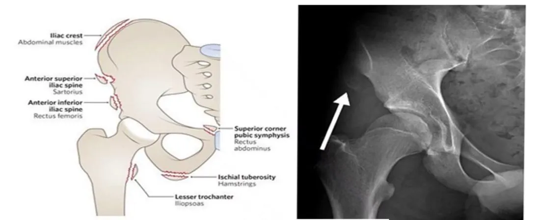
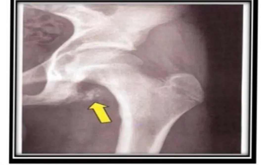
Avulsion fracture of the ischial tuberosity
- Sudden acute pain with a sensation of popping or tearing during a rapid hip flexion movement, and an inability to Weight-bearingPutting body weight through the leg or foot.General meaning, not from the slides.
- Bruising and swelling develop over the following few hours and days, with weakness and pain on prone hip extension, and possibly a visible and PalpationExamining by feeling with the hands.General meaning, not from the slides deformity of the hamstrings.
- Management is usually conservative: offloading the hamstrings with crutches and gentle active and passive movements to avoid joint stiffness.
- Once able to weight-bear, a progressive strengthening and return to play program can commence.
- If the bony fragment is displaced more than 2.5 cm, surgery may be recommended to restore congruity of the tendon and ensure return to sport.
General rehabilitation of hip injuries in sport
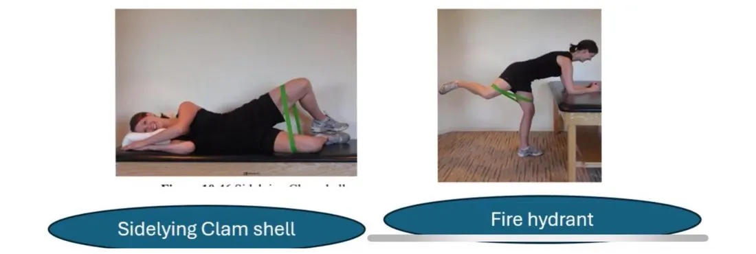
Three components: 1. Hip mobility and flexibility exercise, 2. Gluteus muscle activation and strengthening (the Powers ProgramThe slides' program of eight phases for gluteus maximus activation, strengthening and function, with an emphasis on triplanar GMax function.), 3. Advanced functional training.
1. Hip mobility and flexibility exercise
- Hip mobility can be restored through passive and active manual therapy.
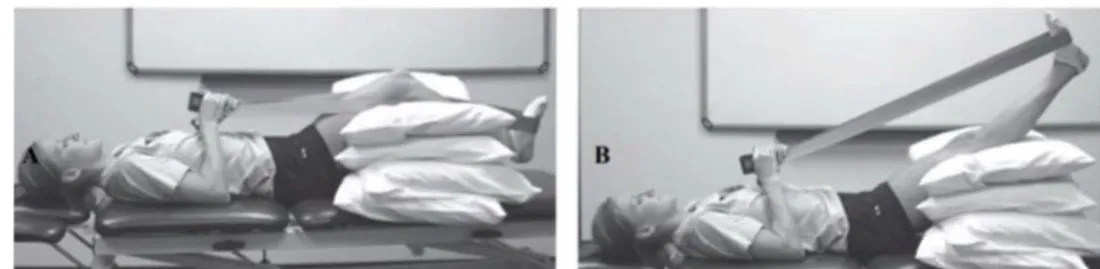
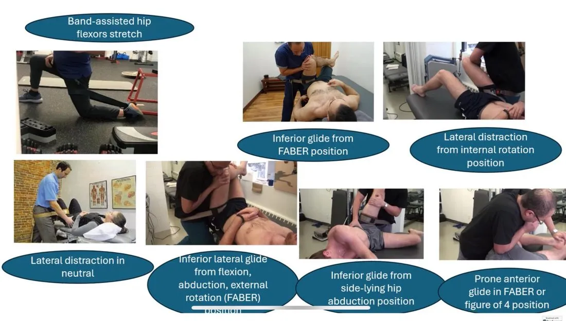
- One of the most effective ways is for the therapist to mobilize the hip joint using a Mobilization beltA strap used by the therapist to apply a gentle pulling or gliding force to the hip joint.General meaning, not from the slides to “unlock” a stiff hip in different directions.
- Care should be taken not to overdo the mobilization and to make sure it is the correct treatment of choice. The therapist should avoid belt mobilization for patients with unstable conditions or after joint replacement surgeries.
- These techniques are good for patients with mechanical “locking” of the joint or for patients with Loose bodiesSmall fragments of bone or cartilage floating free inside a joint.General meaning, not from the slides and Femoroacetabular impingementAn abnormal shape of the proximal femur and acetabulum that raises contact forces in hip flexion, potentially injuring the labrum and cartilage and causing pain..
Techniques pictured on slide 68: band-assisted Hip flexorsThe muscles at the front of the hip that lift the thigh toward the trunk.General meaning, not from the slides stretch; lateral distraction in neutral; inferior lateral glide from flexion, abduction, external rotation (FABERFlexion, abduction and external rotation of the hip, also called the Patrick test.General meaning, not from the slides) position; inferior glide from FABER position; lateral distraction from internal rotation position; inferior glide from side-lying hip abduction position; prone anterior glide in FABER or figure of 4 position.
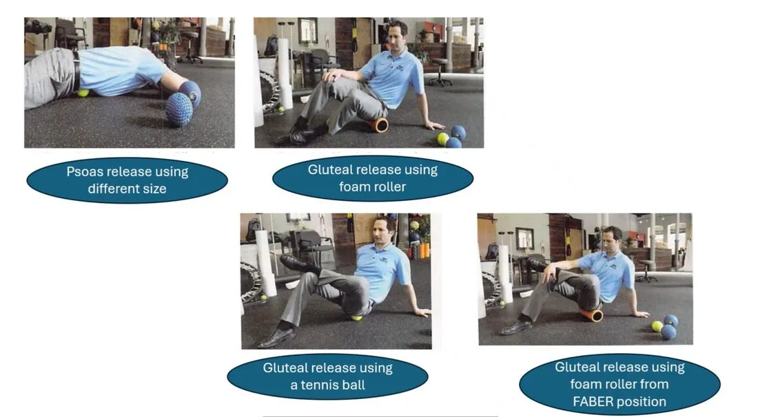
Soft-tissue release pictured on slide 69: PsoasA deep muscle from the lower spine to the top of the femur that flexes the hip.General meaning, not from the slides release using different size (the slide’s wording), gluteal release using a Foam rollerA firm foam cylinder used to roll over muscles for soft-tissue release.General meaning, not from the slides, gluteal release using a tennis ball, gluteal release using a foam roller from FABER position.
2. Gluteus muscle activation and strengthening: the Powers Program
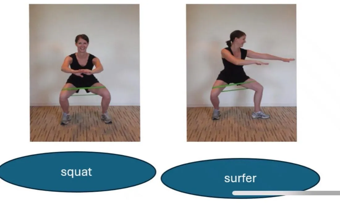
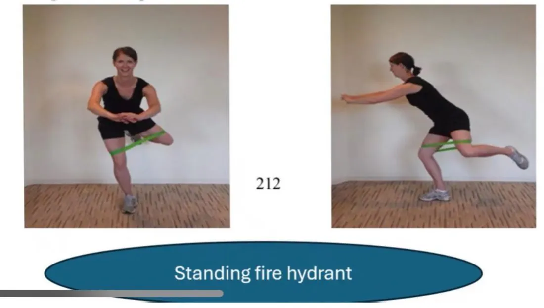
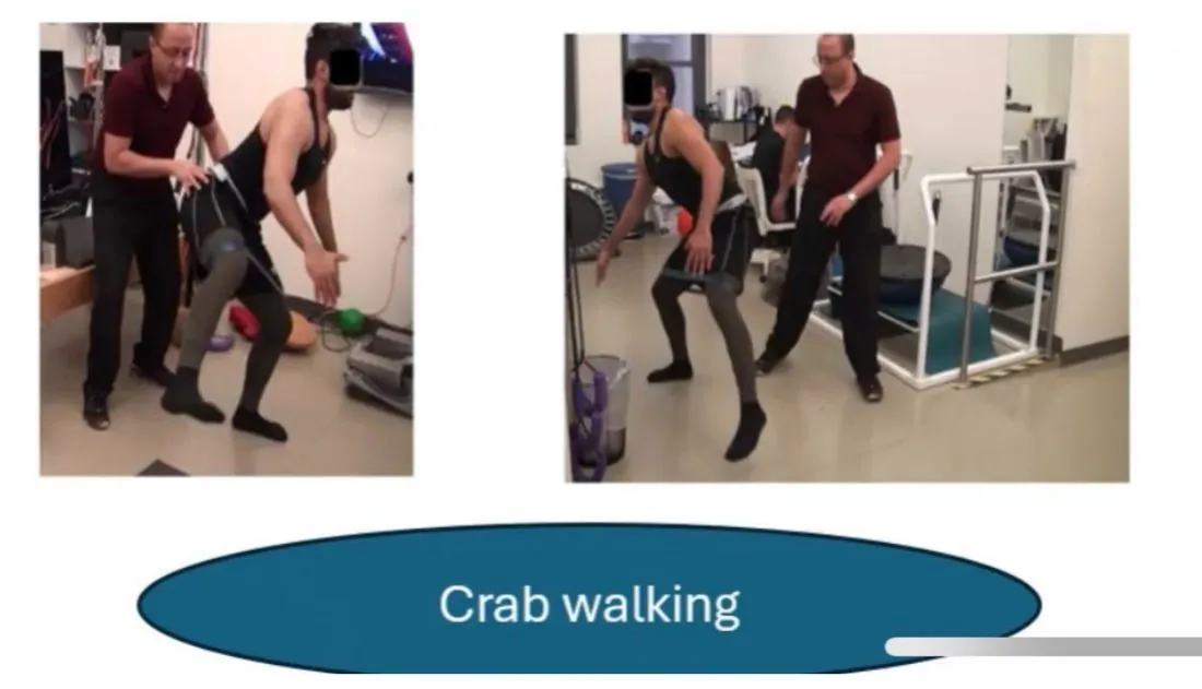
- The program consists of eight phases, with an emphasis on TriplanarWorking in all three planes of movement.General meaning, not from the slides Gluteus maximusThe largest buttock muscle, which extends the hip and rotates it outward.General meaning, not from the slides (GMax) function: abduction, extension, external rotation.
- Phases 1–3: focused on GMax activation.
- Phases 4–5: focused on GMax strength.
- The final three phases emphasize functional applications of the GMax during BallisticFast, explosive movements.General meaning, not from the slides tasks.
- The patient MUST attain the goal repetitions (and holds when required) at one level before progressing to the next level.
| Level | Type | Goal | How often | Exercises |
|---|---|---|---|---|
| 1 | Activation | Hold the end position 60 seconds, 5 repetitions, blue band. May start by holding 5–10 seconds, 5 repetitions, with a yellow, then red, then blue band | Every day | Sidelying Clam shellLying on your side with the knees bent, opening the top knee while the feet stay together.General meaning, not from the slides, Fire hydrantOn hands and knees, lifting the bent knee out to the side.General meaning, not from the slides |
| 2 | Activation | Hold the end position 60 seconds, 5 repetitions, blue band | not stated on the slide | SquatBending the hips and knees to lower the body, then standing back up.General meaning, not from the slides, surfer |
| 3 | Activation | Hold the end position 30 seconds, 5 times, blue band | Every other day | Standing fire hydrant |
| 4 | Strength | 1–2 sets of 15 repetitions with a blue band (except the forward LungeA step forward (or in another direction) with a deep bend of the front knee and hip.General meaning, not from the slides) | Every other day | The same squat as level 2 (different goal), Crab walkingWalking sideways in a low squat, usually with a resistance band around the legs.General meaning, not from the slides, forward lunge |
| 6, 7, 8 | Advanced functional, sport-specific and agility training | Forward and lateral jumping, hopping, single leg jumping and CuttingMaking a sudden change of direction while running.General meaning, not from the slides exercises | — | — |
The slides call these phases (slide 70) and then levels (slides 71–75). Level 5 has no slide of its own. Slide 70 puts it with the strength phases (phases 4–5).
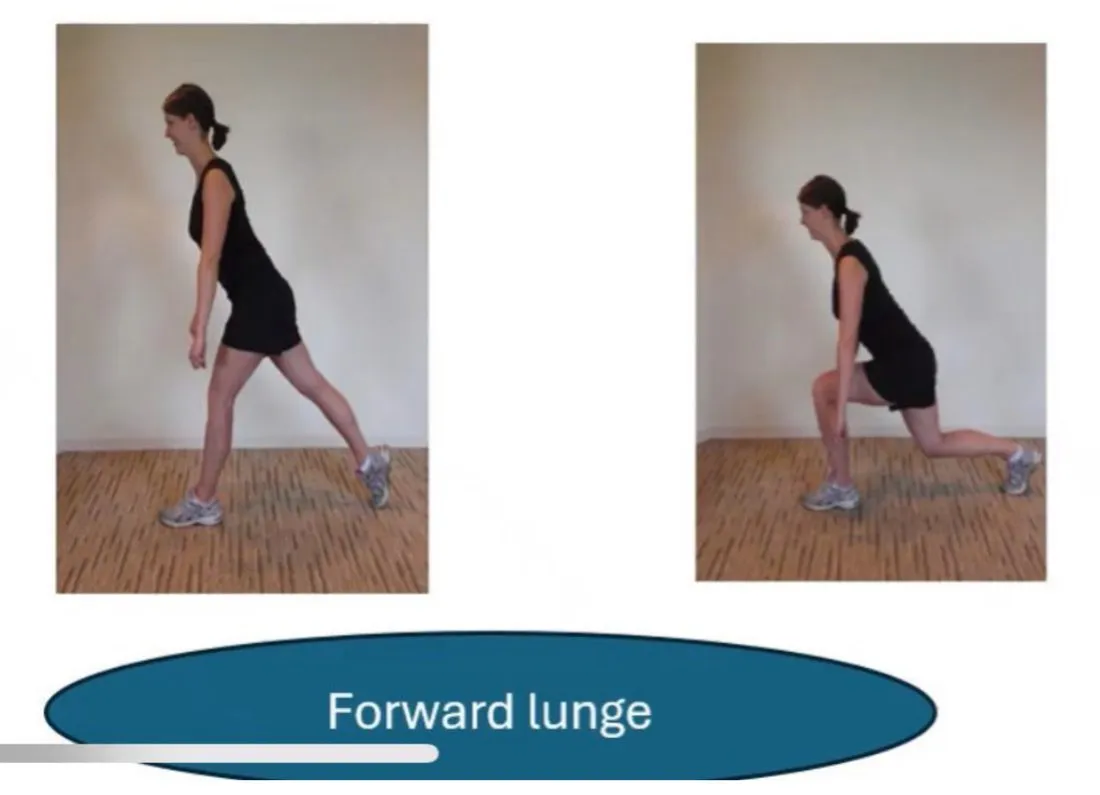
- Levels 6–8 can be a sport-specific training incorporated at the end of the rehabilitation program. The slide says this will be discussed in the next section.
- The final stage of rehabilitation also involves advanced functional training, which activates the upper and lower Kinetic chainA linked series of joints and muscles that work together to produce a movement.General meaning, not from the slides along with core strengthening.
3. Advanced functional training
- The therapist should not attempt this final stage until the previous stages have been completed.
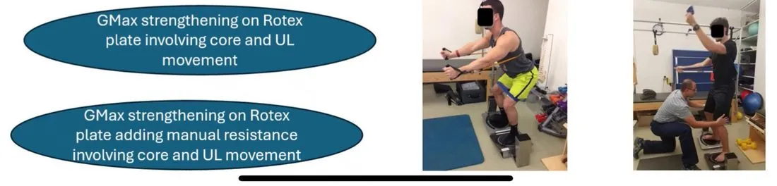
- The reason: these exercises have high demands and involve the upper and lower extremities and the core. If not performed properly or if performed prematurely, injuries can happen and will set the whole rehabilitation back.
- Examples: GMax strengthening on a Rotex plate involving core and ULUpper limb: the arm.General meaning, not from the slides movement, and the same adding manual resistance.
Slide 77 shows further advanced functional training exercises as photos only.
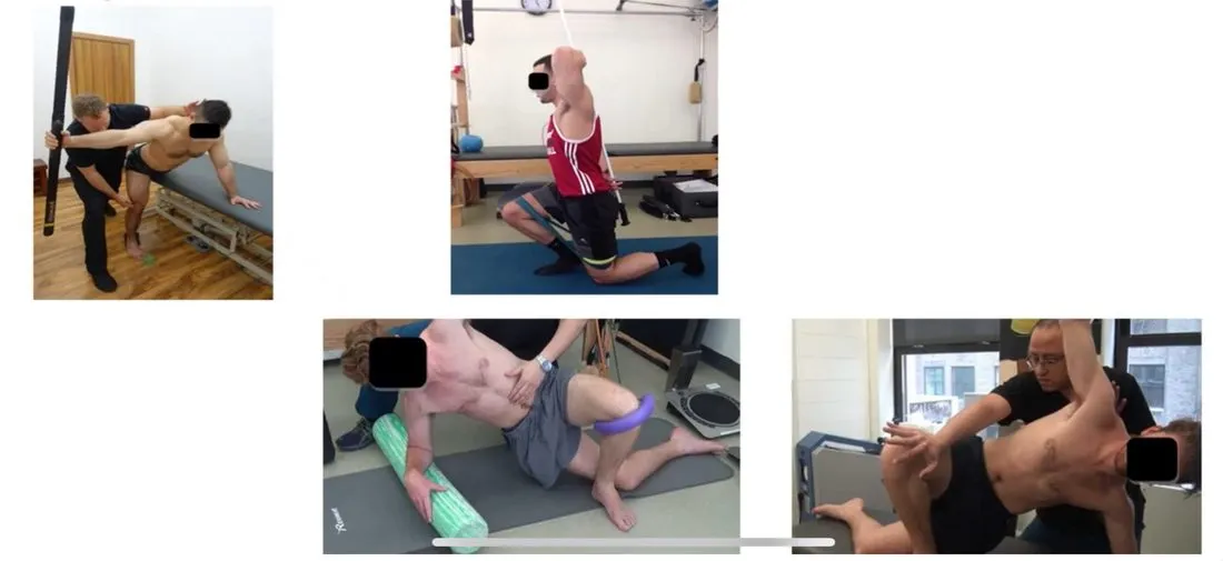
Reminder from slide 76: do not start this final stage until the previous stages are completed, because the exercises make high demands on the upper and lower extremities and the core.